All posts by plancher

exercising a sprained ankle

Effective Strategies for Exercising a Sprained Ankle Safely

Are you looking to exercise your sprained ankle without risking further damage? This guide provides the essential steps for exercising a sprained ankle to support a safe and effective recovery. Learn the precise movements and strategies that help restore strength and flexibility, starting with the most gentle exercises and advancing as your ankle heals. Begin your journey to recovery with confidence, backed by expert advice and structured progression.

Key Takeaways

  • Ankle sprains range from minor ligament stretching to severe high-grade sprains; recovery strategies must be tailored to the specific type and severity of the sprain.
  • Early recovery should employ the RICE method and gradually introduce weight bearing and movements while avoiding prolonged immobilization to prevent secondary damage.
  • Rehabilitation involves gentle range of motion exercises followed by progressive strengthening, balance and proprioception workouts, and advanced functional training, with supportive gear to enhance stability and reduce reinjury risk.
person experiencing pain from ankle sprain

Understanding Your Sprained Ankle

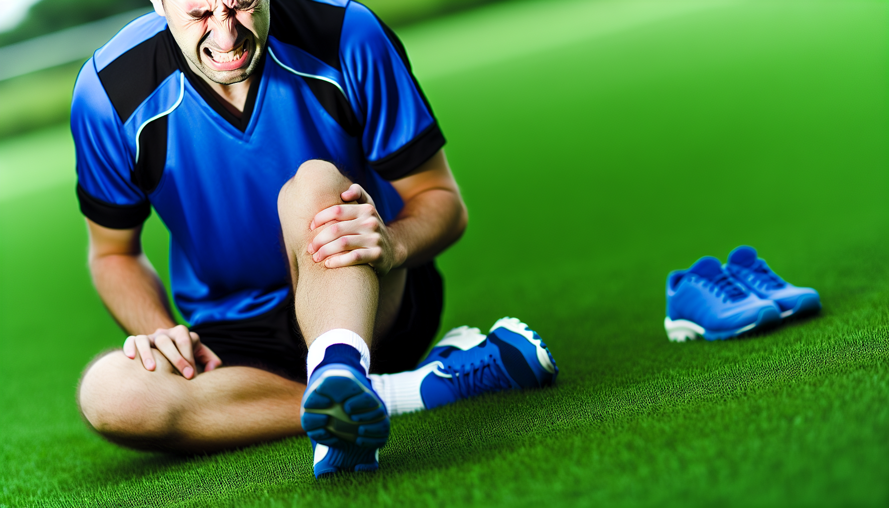
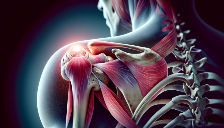
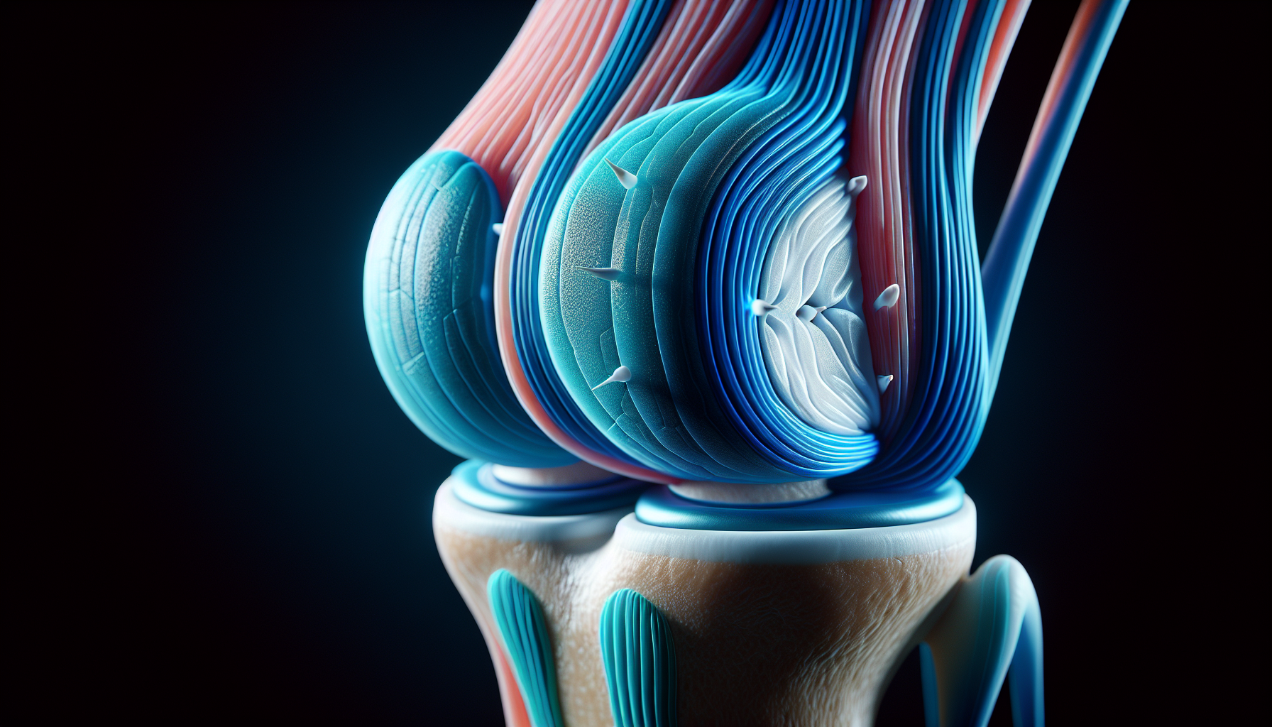
An ankle sprain, a common injury often linked to sports activities, involves damage to the ligaments that support the ankle joint. This complex joint is composed of three bones: the tibia, fibula, and talus. These bones are held together and supported by three main ligamentous systems: the lateral ligament complex, medial deltoid ligament, and syndesmotic ligaments. An injured leg, particularly an ankle sprain, requires proper care and treatment to ensure a full recovery.

Ankle sprains are often caused by the inversion of the foot, which puts stress on the lateral ligament complex, particularly the anterior talofibular ligament (ATFL). The medial deltoid ligament, the strongest of the ankle ligaments, is usually injured during eversion injuries. The type and severity of an ankle injury, or even an injured foot, can vary depending on the position of the foot and the type of rotational force applied.

Ankle sprain severities range from low-grade, involving ligament stretching or minor tears, to high-grade sprains that may include injury to the syndesmotic ligaments. Understanding the specifics of the injured ankle is crucial for determining an appropriate rehabilitation approach, especially in cases of an acute ankle sprain. Syndesmotic injuries, also referred to as ‘high ankle sprains’, are less common and typically affect competitive athletes.

Initial Steps to Exercise Post-Sprain

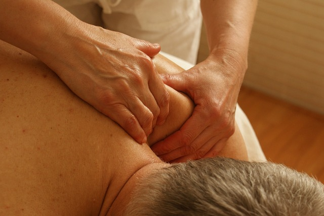
The initial steps post-sprain are crucial for effective recovery. The RICE method, which stands for Rest, Ice, Compression, and Elevation, is a recommended approach to reduce swelling and manage pain. Once immediate care has been addressed, rehabilitation should start promptly, focusing on reducing swelling, pain, and inflammation while protecting the joint from secondary damage due to excessive early exercises.

Gradual weight-bearing and comfortable movements, like supported walking for minor sprains, should be introduced within the first week, aligning with Plancher’s advice to foster healing. The common mistake of prolonged immobilization should be avoided by applying controlled stress to the injured area.

Protecting the ankle during the early stages is crucial, with later stages incorporating careful stress under supervision to aid the recovery process. Rehabilitation exercises should be tailored to the individual’s specific condition, following a doctor’s advice.

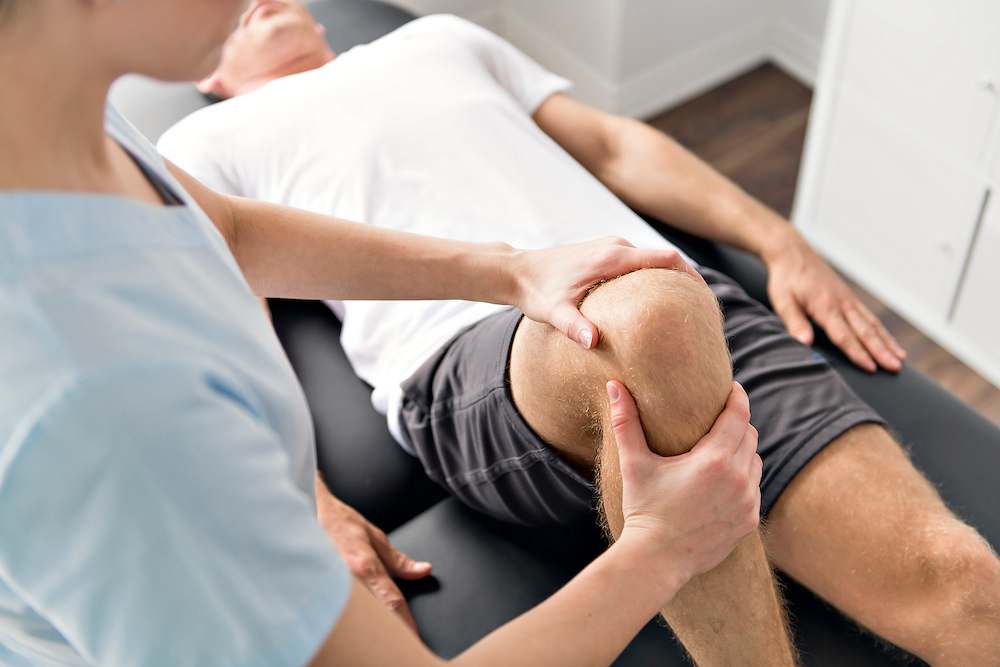
Gentle Range of Motion Exercises

Once the initial care phase has been navigated successfully, it’s time to begin gentle range of motion exercises, focusing on knee motion. To begin these exercises, support the leg while allowing the foot and ankle to hang freely, typically done by performing a knee motion sit and positioning the leg on another chair.

Engage in a series of exercises to promote a gentle range of motion in the sprained ankle. These exercises include:

  • ‘Flex’
  • ‘Extend’
  • Inward and outward bends
  • Ankle circles
  • Foot alphabet exercises

Perform each exercise by holding each position for 10 to 15 seconds and repeating the movements 10 times, twice a day.

These gentle range of motion exercises aid in regaining mobility and strengthening the muscles around the ankle, helping to prevent chronic ankle instability without causing excessive strain. Remember, the pace should be guided by pain, and any movement that causes increased pain should be avoided.

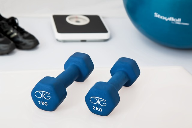
Progressive Strengthening Techniques

Progressive strengthening is a key step in the recovery process, encouraging the building of strength over time. Towel curls are recommended as the first of the strengthening exercises after a normal range of motion returns in a healing ankle.

Towel curls are performed by sitting and using the toes to pull and push a towel on the floor. This can be repeated 3 to 5 times a day to strengthen foot muscles. To stimulate and strengthen the plantar fascia, use a golf ball to gently roll under the foot’s arch and pick up marbles with the toes to condition the plantar flexors.

Once these exercises are mastered, progress to isometric exercises, such as pushing the foot against a sturdy object. Then, introduce elastic stretch band exercises for dorsiflexion and plantar flexion to further strengthen the anterior tibialis and gastrocnemius-soleus complex.

Flexibility and Calf Muscle Care

A key component of ankle sprain recovery is maintaining flexibility and caring for your calf muscles. Regular stretching should be done daily, especially before and after any physical activity, to prevent stiffness and reinjury of the calf muscles.

Seated and standing calf stretches enhance the flexibility of the calf muscles during recovery from a sprained ankle. These can be performed with feet shoulder-width apart, toes forward, back straight, shoulders back and down, raising the heels, with extended but not locked knees.

To perform a seated calf stretch, sit with a straight back and core engaged, place feet flat on the ground, and press down on the balls of the feet, lifting the heels. Using a step or stair for calf raises can enhance the stretching range of motion, allowing for a deeper stretch.

Begin stretching the Achilles tendon when it is possible to do so without pain.

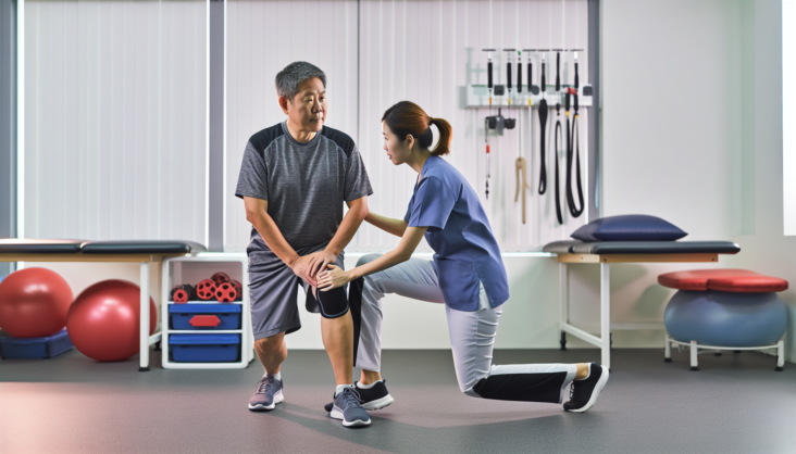
Balance and Proprioception Workouts

Balance and proprioception workouts also play a crucial role in ankle sprain recovery. These exercises should start when a patient can stand without pain, provided they were steady on their feet before the injury.

Establishing proprioception and ankle stability can begin with:

  • Single-leg balances near a support surface
  • Balancing on a wobble board
  • Using an ankle disk
  • Balancing on one leg with the ankle inverted

Ankle rhythmic stabilization is a technique aimed to improve balance and control as part of a comprehensive ankle rehabilitation process. As rehabilitation progresses, individuals may start with simpler variations of balancing tasks and gradually introduce more challenging variations as their strength and stability improve.

Assessment of postural stability is essential during the rehabilitation process, providing valuable feedback for progress and exercise adjustment.

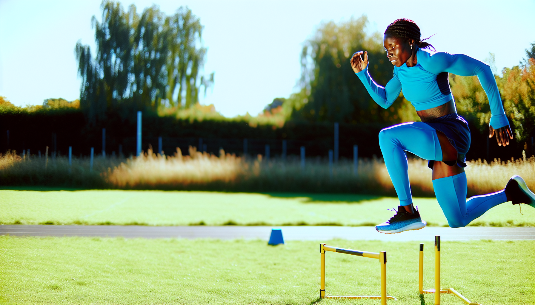
Advanced Functional Training

Advanced functional training is a step up from basic exercises, providing a more dynamic approach to recovery. Heel raises, for example, enhance calf muscle strength by lifting the heel as high as possible while keeping the knee straight. Heel raises are performed by standing behind a chair for support, rising onto the toes, and gradually lowering back down without favoring the injured side. Once basic heel raises are mastered, a squat variation that targets calves and dynamic ankle stability can be integrated into the exercise regime.

The Y-Balance exercise is a challenging movement that enhances balance, proprioception, and strength by moving the ankle through variable positions. Forward and lateral BOSU lunges combined with single-leg balancing enhance dynamic stability and confidence in the rehabilitated ankle.

Lateral hop and stick exercises are useful in enabling control of lateral movements, and vital for regained stability in the ankle post-sprain.

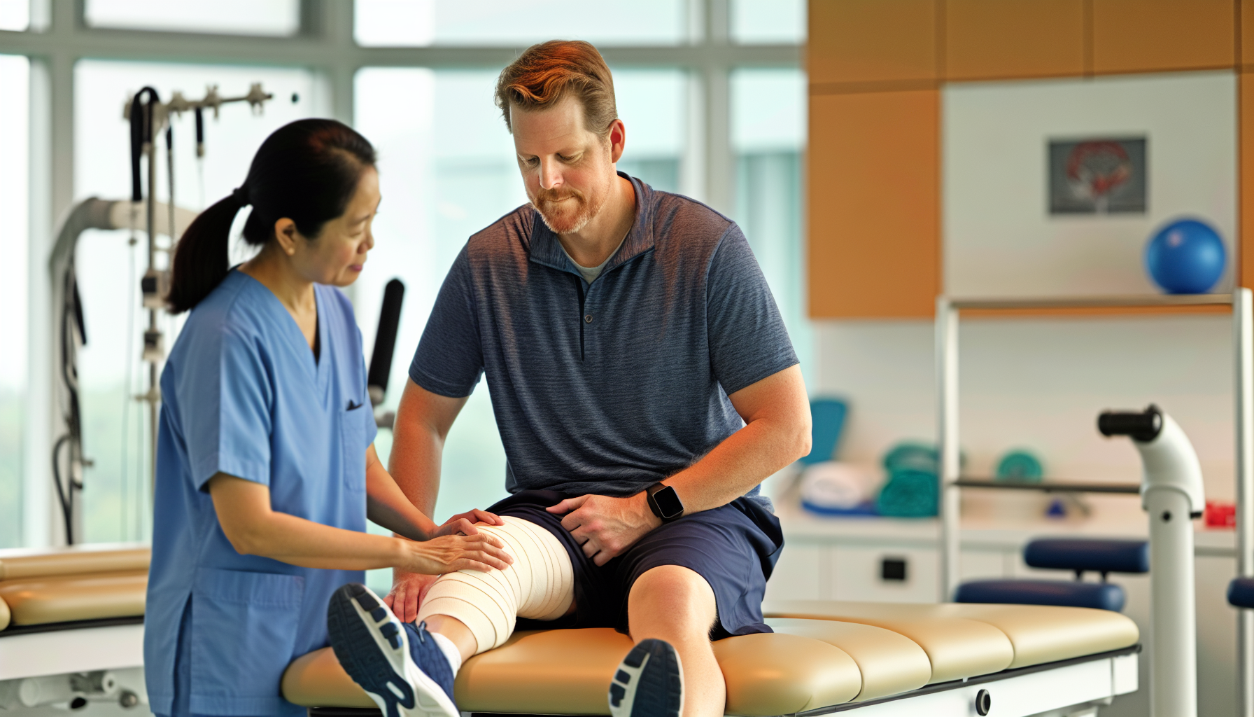
Utilizing Supportive Gear

Supportive gear, such as ankle braces and taping, can provide additional stability during the recovery process. Ankle bracing during the early stages of rehabilitation can improve proprioceptive feedback and potentially reduce the risk of re-injury.

The lateral ankle stability tape technique can provide feedback on ankle position and improve proprioception after a sprain. It’s essential to choose ankle braces based on the user’s shoe size or ankle circumference to ensure the proper fit and support.

There are various types of ankle braces, each providing a different level of support and comfort. Lace-up ankle braces allow users to adjust tightness for maximum support and a customized fit. Neoprene material in ankle braces provides both support and heat insulation, beneficial for pain, but may not be suitable for all-day use due to lack of breathability.

Fabric and knit ankle braces offer better breathability, keeping the feet cooler, and might be preferable for extended wear. Strap-enhanced ankle supports offer advanced protection by allowing customizable compression levels and are typically available in figure 8 or figure 6 configurations.

Reinforcing Safe Practices

Safe practices are paramount in the recovery process. Pain should guide the pace of rehabilitation exercises for a sprained ankle. It’s crucial to listen to the body’s response to injury, which should dictate the rehabilitation approach for future function and athletic performance.

A personalized rehabilitation program should be constructed based on empirical evidence and knowledge from structured rehabilitation protocols. Kevin D. Plancher advises a cautious approach to recovery, emphasizing the importance of a gradual return to normal activities. The focus should always be on safety and effective recovery.

Recovery Tips from a Sports Medicine Expert

Today’s guide to ankle sprain recovery is informed by the expertise of Kevin D. Plancher, an orthopaedic surgeon and sports medicine expert. With his extensive medical education including a Doctorate in Medicine and a Master’s in Science in Physiology, complemented by an MPH, Kevin D. Plancher’s clinical insight is backed by a strong academic foundation.

His roles as a Clinical Professor at Montefiore Medical Center/Albert Einstein College of Medicine and an Adjunct Clinical Assistant Professor at Weill Cornell Medical College, Cornell University signify his standing in the medical community. Through his leadership at Plancher Orthopaedics & Sports Medicine and his establishment of the non-profit Orthopaedic Foundation for Active Lifestyles, Kevin D. Plancher has extended his impact to the broader community, promoting health and recovery.

Summary

Recovering from an ankle sprain is a journey, one that requires understanding, patience, and above all, guided exercises. By understanding the specifics of your injury, adhering to a personalized recovery program, and following expert advice, you can navigate this path to recovery effectively and safely. Remember, the journey is not a sprint but a marathon, and each step you take brings you closer to your goal. Contact Plancher Orthopaedics to see how we can further help you with your sprained ankle!

Frequently Asked Questions

What is the first step in ankle sprain recovery?

The first step in ankle sprain recovery is to follow the RICE method – Rest, Ice, Compression, and Elevation – to reduce swelling and manage pain effectively.

What are some examples of gentle range of motion exercises?

Some examples of gentle range of motion exercises include flexing, extending, inward and outward bends, ankle circles, and foot alphabet exercises. These exercises can help improve flexibility and reduce stiffness.

How can supportive gear aid in recovery?

Supportive gear such as ankle braces and taping can improve stability, enhance proprioceptive feedback, and reduce the risk of re-injury, aiding in the recovery process.

What is the role of balance and proprioception workouts?

Balance and proprioception workouts play a crucial role in improving stability and control, which are essential for effective ankle sprain recovery.

Who is Kevin D. Plancher?

Kevin D. Plancher is an orthopaedic surgeon and sports medicine expert specializing in knee, shoulder, elbow, and hand injuries. He is a renowned professional in his field.

Read More
pain after knee replacement surgery

Pain relief after knee replacement surgery

Pain after knee replacement surgery is a typical part of the healing process. But what causes it, and how can you manage it effectively? This article directly addresses these pressing concerns with simple explanations and evidence-based pain relief strategies to foster smoother recovery for patients grappling with this common post-surgical issue.

Key Takeaways

  • Effective post-surgery pain management in knee replacement is crucial for a quicker recovery process, involving medication, physical therapy, and alternative methods such as ice application and elevation.
  • Postoperative knee pain can result from various causes including infection, implant issues, or nerve damage, and addressing these issues promptly is key to effective pain alleviation and optimal recovery.
  • Maintaining a healthy weight, making home safety modifications, engaging in suitable activities, and having a support system are vital strategies for ensuring a smooth recovery from knee replacement surgery.

Pain Management After Knee Replacement Surgery

Person undergoing physical therapy after knee replacement surgery
Patient at the physiotherapy doing physical exercises with his therapist

Successfully controlling pain after knee replacement surgery can accelerate the recovery process, including hastening physical therapy sessions, diminishing inflammation and promoting better healing. For superior pain control, strategies for managing discomfort might include a combination of medications such as opioids and non-steroidal anti-inflammatory drugs (NSAIDs), along with patient-administered analgesia pumps and nerve blocks. Although experiencing pain for several weeks post-replacement surgery is expected, proper management of this discomfort can enhance prospects for a more successful recovery.

Enhancing methods of pain relief following knee replacement procedures can contribute to reduced duration of hospital stays and decrease associated expenses while notably improving patient contentment and quality of life. Subsequent sections will delve into diverse tactics employed in addressing post-surgery pain starting from pharmaceutical interventions aimed at alleviating suffering.

Medications for Pain Relief

Following knee replacement surgery, it is common for individuals to be prescribed a range of medications such as NSAIDs, acetaminophen, and opioids to manage postoperative pain. Specifically, narcotic pain medications like morphine, hydromorphone, hydrocodone, oxycodone, and meperidine play a crucial role in immediate post-surgical pain relief. These drugs must be taken strictly according to medical advice and their use tapered off as the level of pain subsides.

Continuous supervision of patients’ respiratory functions and oxygen levels becomes necessary when they’re on opioid treatments due to the risk of respiratory depression associated with these drugs. Following proper dosage guidance for taking this medication can contribute significantly towards minimizing swelling, which facilitates physical therapy endeavors while also accelerating recovery following knee replacement surgeries.

To conventional medicines mentioned earlier, there are alternative options such as liposomal bupivacaine available. This drug can be administered directly to the site where surgery was performed, providing sustained relief from discomfort over approximately three days following the procedure. Patients might receive other types of medications including ondansetron or promethazine intended specifically for counteracting adverse reactions arising out either from anesthesia or narcotics employed during their comprehensive approach towards managing post-operative pains after undergoing Knee Replacement procedures.

Physical Therapy and Pain Reduction

Beginning just a few hours after knee replacement surgery, engaging in physical therapy plays an instrumental role in decreasing postoperative pain and aiding the recovery process. This is achieved through immediate mobilization and exercises designed to strengthen muscles. The rehabilitation plan structured by physical therapy focuses on enhancing flexibility, muscle building, and fulfilling objectives specific to each patient’s needs.

A comprehensive regimen of physical therapy after knee replacement encompasses activities starting from within the hospital setting to home-based routines as well as sessions with outpatient therapists. The key targets of this full-scale program include:

  • Mitigating pain
  • Improving mobility
  • Restoring optimal functionality of the knee
  • Professional guidance from a dedicated physical therapist
  • Following a customized rehabilitation strategy
  • Re-establishing movement capabilities
  • Knee fortification

All these efforts are directed toward alleviating post-surgical discomfort via safe yet effective therapeutic exercises.

Alternative Pain Relief Methods

Alternative methods for alleviating postoperative knee pain can greatly enhance pain relief efforts following a knee replacement. The application of ice serves as an effective method to control pain after surgery, offering a significant reduction in discomfort both on its own and in combination with medications. It’s advised to apply ice packs no longer than 20 minutes at one time to help reduce swelling.

Incorporating leg elevation and ensuring sufficient rest are also vital components of an effective strategy for dealing with post-knee replacement surgery discomfort. Employing techniques like guided imagery, biofeedback, and cognitive-behavioral therapy could be beneficial in coping with pain experienced after undergoing knee replacement. Alternative approaches such as acupuncture have been known not only to alleviate knee-related pains but also to diminish swelling and promote enhancements in range of motion between the first week up until three weeks following the replacement procedure.

Potential Causes of Postoperative Pain

Understanding the underlying causes of postoperative pain is essential for its successful management and reduction. Persistent pain following total knee arthroplasty (TKA) has complex origins that include biological, surgical, and psychological factors.

Key contributors to postoperative discomfort may consist of:

  • Levels of pre-surgery pain which can predict how intense the post-surgery pain might be
  • The patient’s age since older individuals could face heightened levels of discomfort after surgery
  • Mental states like anxiety and depression that have been known to amplify perceived pain during the recovery process

Gaining insight into these elements allows medical professionals to formulate appropriate strategies aimed at managing and mitigating pains particularly associated with postsurgical conditions.

Identified risk determinants for long-standing chronic agony following TKA include:

  • Intensity of pre-procedure suffering
  • Subpar mental health status
  • A propensity towards magnifying one’s perception of painfulness
  • Concurrent additional medical issues

Subsequent sections will explore in greater detail certain specific sources behind lingering pains after surgery such as infections, complications related directly to the implant itself, or injuries inflicted upon nerves.

Infection

Following knee replacement surgery, infections can trigger pain by the secretion of inflammatory molecules like IL-1. Common culprits include bacteria such as Staphylococcus aureus and Corynebacterium. Indicators that an infection may have set in are:

  • Escalating pain with movement or at rest
  • Altered appearance of the surgical site
  • Experiencing chills
  • Developing a high fever

It’s critical to obtain medical attention promptly if these symptoms emerge.

Not only do these infections cause discomfort, but they can also extend the duration of the recovery process after knee replacement surgery. Ensuring cleanliness and sterility during post-operative care is essential. Alerting your healthcare providers about any potential signs of infection is crucial for timely intervention, helping to avoid added complications and facilitating a smoother path to recovery from your knee replacement.

Implant Issues

Issues with the implant can lead to discomfort following a knee replacement surgery. Over time, surfaces of the implant may wear out and components might become loose, leading to pain. The loosening of the total knee arthroplasty implant is commonly linked to failure due to factors like instability, improper positioning of components, or bone loss caused by polyethylene wear.

Should patients notice that their knee feels unstable or seems to collapse unexpectedly after total knee replacement surgery, it could indicate a problem with the implant’s security. In such cases where there are undeniable indications for revision surgery because of these issues related directly to the prosthesis used in total knee arthroplasties (TKA), discussion on another operation becomes necessary. Revision procedures for persistent postoperative pain after TKA are complex. They entail removing existing prostheses potentially adding bone grafts and fitting specialized new implants. This route should be reserved when definitive causes tied specifically back to complications from initial replacement surgeries can be confirmed since results typically do not favor those undergoing revisions solely based on undiagnosed chronic pain reasons post-total joint replacement operations.

Nerve Damage

Nerve damage incurred during knee replacement surgery is another potential source of persistent postoperative pain, and it can manifest as ongoing severe discomfort, a loss of sensation, or muscle weakness in the limb that was operated on. For example, if the saphenous nerve’s infrapatellar branch is harmed during a knee replacement procedure, it could result in the development of an agonizing neuroma and adversely affect the function of the knee.

Persistent medial pain in the knee area specifically associated with an infrapatellar saphenous nerve neuroma may be evident when other explanations are ruled out. The routine incision made for total knee arthroplasty often cuts through this particular nerve branch which might then lead to forming a painful neuroma due to its entrapment within scar tissue following surgery. Although employed during operations to mitigate discomfort, nerve blocks may inadvertently impair muscular activity in the lower extremity. This presents a significant problem since actions such as walking and engaging in physical therapy are essential both for alleviating pain and facilitating recuperation after undergoing total knee replacement surgery.

Monitoring and Reporting Pain

Monitoring and communicating about post-surgical pain is an essential element of aftercare following a knee replacement. Utilizing a scale from 1 to 10, patients are expected to self-evaluate their discomfort levels, which serves as a critical tool for gauging the success of pain relief efforts following knee replacement surgery. To adequately address any issues with pain management, reassessments should be performed routinely.

It’s important that open communication is maintained with your surgeon and that you set realistic expectations for recuperation after undergoing a knee replacement procedure. Consider adopting these measures.

  1. Undergo thorough evaluations to fully grasp both the nature and severity of your post-operative pain.
  2. If persistent or unusual discomfort occurs outside the anticipated recovery timeframe, promptly discuss this with your healthcare provider.
  3. Adhere strictly to medical guidance provided by your surgeon concerning additional therapeutic options or amendments needed in your healing protocol.

Adhering closely to these recommendations will help facilitate more effective convalescence following knee replacement surgery and improve overall outcomes related to managing postsurgical pain.

Addressing Persistent Pain

Chronic knee pain, defined as discomfort lasting more than three months after a total knee arthroplasty (TKA), can significantly affect an individual’s quality of life-related to health. Around 20% of patients who undergo total knee replacement remain unsatisfied with the outcome, frequently due to persistent and severe knee pain.

In order to address continuous pain following TKA, several strategies may be employed. The premier approach for managing periprosthetic joint infection post-TKA is two-stage revision arthroplasty incorporating an antibiotic spacer along with thorough debridement and antibiotics selected according to the specific infection.

Complications from knee revision surgery are acknowledged concerns and include potential nerve or blood vessel damage as well as risks associated with blood clots. These issues can be effectively reduced by employing meticulous surgical techniques complemented by appropriate antibiotic use.

Tips for a Smooth Recovery

Achieving a successful recovery following knee replacement surgery requires careful planning and dedication to certain key practices, even before the surgical procedure. Ensuring that you keep your weight in check after a knee replacement is critical, as it helps reduce undue stress on the healing joint and diminishes the risk of causing injury or damage to the other knee.

Research indicates that within two years post-surgery, 66% of individuals who have undergone a knee replacement tend to gain approximately 15 pounds. This statistic underscores the importance of incorporating weight management strategies into one’s postoperative regimen. Here are several actionable suggestions for managing weight during this time.

  • Embrace a nutritious diet filled with high-fiber items, proteins low in fat content, and carbohydrates with complex structures.
  • Consume water before eating meals which can aid in achieving satiety sooner.
  • Practice mindful consumption by eating at unhurried paces.
  • Organize your meal preparation before undergoing surgery by creating healthy dishes ahead of time so they’re ready when needed during your convalescence.

Implementing these guidelines into your daily routine after having undergone replacement surgery for your knees can be instrumental in helping you retain an optimal body mass thus facilitating smoother progress through each phase of recuperation.

Home Modifications

Home modifications for safe recovery

Preparing your living space for a safe and manageable recovery is an essential part of the process before undergoing knee replacement surgery. To do this, you may:

  1. Clear out any clutter from your home to eliminate potential tripping hazards like loose rugs, stray furniture pieces, and electrical wires.
  2. If possible, add ramps in place of steps which can help simplify mobility around the house.
  3. Apply non-slip treatments to floors that tend to be slick to prevent falls.

Implementing these changes will help ensure a secure environment conducive for recuperation after your knee replacement.

It’s also advisable to set up a recovery area on the ground level of your home if available, as this circumvents the need for stair navigation post-surgery. Within this designated area, make sure that daily necessities such as eyewear, electronic gadgets along with their chargers, reading material, snacks, and drinks are easily reachable without straining yourself. Moreover, having grab bars by shower areas or toilets can significantly increase safety during bathroom use. Utilizing elevated toilet seats alongside having a seat while showering can Facilitate comfort and stability following knee replacement surgery.

Activity Recommendations

It is imperative to maintain an active lifestyle following knee replacement surgery. It’s just as vital to partake in activities that don’t overburden the freshly implanted knee. You can start driving again 4 to 6 weeks after undergoing replacement surgery once healing has sufficiently progressed and leg movement control has been re-established.

Initially, post-surgery mobility should involve using walkers or crutches, while avoiding stairs until a surgeon or physical therapist approves such activity. To stay active yet protect the new knee joint from unnecessary stress, participating in low-impact sports like swimming, cycling, and golf is advisable. Always seek advice from your surgeon before starting any exercise regimen or athletic endeavors after the surgery.

Support System

A strong and reliable network of support is essential for a smooth recovery following knee replacement surgery. During the recovery period from this surgery, patients often require assistance with various daily activities including:

  • meal preparation
  • running errands
  • personal hygiene care
  • managing laundry

This is due to their reduced ability to move around freely. Before the procedure, social workers or discharge planners can be instrumental in coordinating either at-home aid or arranging for a temporary stay in an extended care facility to ensure the patient’s needs are met.

Emotional support provided by friends, family members, and work associates plays an invaluable role both in deciding to undergo knee replacement and during the postoperative convalescence phase. Having a solid circle of emotional support contributes significantly to sustaining positive spirits and active participation needed for successful rehabilitation after such surgeries.

Long-Term Care and Maintenance

Securing a life devoid of pain after knee replacement surgery is not only about the success of the procedure itself but also hinges on ongoing care and safeguarding the endurance of the prosthetic joint to ensure smooth re-engagement with daily tasks. Keeping scheduled visits with your surgeon post-surgery is critical for tracking the status of your knee replacement as time progresses.

Around 80% remain free from needing any additional surgical intervention two decades following their initial knee replacements due to meticulous long-term upkeep. Much like automobile brake systems, high-impact activities can expedite deterioration in joint prostheses. Hence it’s crucial to participate in low-impact exercises while also managing body weight effectively. Undertaking timely surgery specifically aimed at liner substitution can circumvent more elaborate revision surgeries later on.

Summary

This manual has comprehensively addressed various aspects of managing discomfort following a knee replacement, including the employment of medication and physical therapy, adjustments to living spaces, and the necessity for robust support networks. It also investigated potential origins of pain after surgery such as infections, complications with the prosthetic joint, and nerve injuries while highlighting how vigilant monitoring and communication are key to effective pain control.

The task of controlling discomfort following a knee replacement can seem daunting. Equipped with appropriate techniques, resources, and encouragement, it is indeed feasible to navigate towards a fruitful recuperation reaping benefits from your new knee. Bear in mind that this pathway does not have to be traversed solitarily—assistance is abundant at your disposal. March forward on this journey toward an existence devoid of pain armed with confidence and positivity.

Frequently Asked Questions

How long does it take to walk normally after knee replacement?

Around six weeks following a knee replacement operation, the majority of people are able to ambulate independently without needing any walking aids. Recovery durations can differ.

Usually, the recuperation phase for a knee replacement surgery spans approximately 12 weeks.

What is the hardest part of knee replacement recovery?

During the initial six weeks following a knee replacement, patients often struggle with substantial pain and severe swelling, particularly in the first week, which is considered the most challenging period of recovery.

How do I get my knee to stop hurting after surgery?

To manage discomfort and facilitate the healing process after surgery, consider engaging in light walking exercises, using ice therapy at the site of your surgical incision, and adhering strictly to the prescribed pain medication regimen to mitigate knee pain.

What helps with pain at night after knee replacement?

To mitigate nighttime pain following a knee replacement, administer your medication approximately one hour before bedtime so that it becomes effective by the time you are ready to sleep. Apply ice to your knee just before going to bed and think about propping up your leg on a wedge pillow. This can enhance blood flow.

How long does the pain last after a knee replacement?

Typically, the discomfort experienced following knee replacement surgery subsides gradually over the first two to three weeks as part of the healing process.

Read More

Kevin D. Plancher, MD, MPH, FAOA, FAOS Member of BHS

New York and Greenwich, CT
April 11, 2024

Dr. Kevin Plancher is delighted to announce his membership in the British Hip Society. The British Hip Society (BHS) supports training, education and research to promote the very best care for patients with hip related conditions. The Society works closely with allied professional bodies like the British Orthopaedic Association to advise on standards of care for patients and welcomes hip specialty surgeons, at all career stages, to our membership, including trainees, SAS grades and consultants.

Kevin D. Plancher, MD, MPH, FAOA, FAOS is a board-certified orthopaedic surgeon and founder of Plancher Orthopaedics & Sports Medicine. Dr. Plancher is a Clinical Professor, Department of Orthopaedic Surgery, at Montefiore Medical Center/Albert Einstein College of Medicine and an Adjunct Clinical Assistant Professor of Orthopaedic Surgery, at the Weill Cornell Medical College, Cornell University in New York. Since 2001, he has been listed annually in the Castle Connolly directory as a “top doctor” in his field.

Plancher Orthopaedics & Sports Medicine is a comprehensive orthopaedics and sports medicine practice, specializing in arthroplasty with offices in New York City and Greenwich, CT. Leaders in orthopaedics, sports medicine, and acute emergency treatment of sports injuries and rehabilitation utilizing the latest techniques in orthopaedic care. plancherortho.com

Read More

Kevin D. Plancher, MD, MPH, FAOA, FAOS Ranked Top 1% 

New York and Greenwich, CT
April 11, 2024

Dr. Kevin Plancher is honored to to be named a Hip and Knee Surgery Research All-Star and ranked in the top 1% of surgeons nationwide. This distinction acknowledges the substantial and high-quality research contributions I’ve made through co-authored publications. Curated by Avant-Garde Health, the Healthcare Research All-Star lists feature exclusively the top 5% of hospitals and surgeons who are leading the way in innovative research. Learn more at here.

Kevin D. Plancher, MD, MPH, FAOA, FAOS is a board-certified orthopaedic surgeon and founder of Plancher Orthopaedics & Sports Medicine. Dr. Plancher is a Clinical Professor, Department of Orthopaedic Surgery, at Montefiore Medical Center/Albert Einstein College of Medicine and an Adjunct Clinical Assistant Professor of Orthopaedic Surgery, at the Weill Cornell Medical College, Cornell University in New York. Since 2001, he has been listed annually in the Castle Connolly directory as a “top doctor” in his field.

Plancher Orthopaedics & Sports Medicine is a comprehensive orthopaedics and sports medicine practice, specializing in arthroplasty with offices in New York City and Greenwich, CT. Leaders in orthopaedics, sports medicine, and acute emergency treatment of sports injuries and rehabilitation utilizing the latest techniques in orthopaedic care. plancherortho.com

Read More
ACL Tears

Understanding ACL Tears: Comprehensive Guide to Symptoms, Treatments, and Recovery

Suffering from knee pain or instability? An ACL tear, one of the most common knee injuries, could be the culprit. Understanding ACL tears—their signs, causes, and your path to recovery—is essential. This article provides concise, easy-to-understand information on ACL tears to aid in your diagnosis and decision-making for treatment. Get the facts quickly and start your journey to recovery with confidence.

Key Takeaways

  • ACL tears frequently occur in sports with sudden directional changes and are more common in women due to biomechanical differences, with a significant number of tears being non-contact injuries.
  • Diagnosis of ACL tears involves physical exams and an MRI to confirm injury severity, while treatment options range from non-surgical methods like rest and physical therapy to surgical reconstruction based on injury severity and patient needs.
  • Preventing ACL injuries is essential and can be achieved through training and proper equipment, while sports medicine provides critical care for diagnosis, treatment, and safe return to sports post-ACL injury.

Understanding ACL Tears: Causes and Risk Factors

Active individuals and athletes often suffer from injuries to the anterior cruciate ligament (ACL), including damage to its associated structures such as the medial collateral ligament and lateral collateral ligament. High-impact sports, like basketball, football, soccer, and skiing, tend to report more incidents of ACL tears. Every year in the United States alone, it’s estimated that between 100,000 and 200,000 cases arise, which may necessitate a surgical procedure known as anterior cruciate ligament reconstruction. Notably too is that women are at a greater risk of sustaining an ACL injury compared to men due primarily to differences in biomechanics.

Common Causes

Most ACL tears happen during sports characterized by abrupt halts and directional shifts. The classic scenario leading to an ACL tear involves a sharp twisting motion, with the feet fixed in place while the knees pivot in another direction. This type of serious injury is frequently seen in sports like:

  • soccer
  • basketball
  • skiing
  • football

Such athletic endeavors that require quick pivots or leaping movements are often associated with this form of significant damage.

Risk Factors

Several elements can increase the likelihood of sustaining an ACL injury. Women are known to have a much greater chance of experiencing these injuries than men, especially in sports that require actions such as cutting, pivoting, and leaping. Anatomical characteristics like having increased genu valgum or knock-knees could also boost the risk for ACL injuries. Engaging in contact sports tends to raise the probability of suffering from ACL injuries due to associated movements like turning and valgus stress or hyperextension often resulting from physical collisions.

Intriguingly, around 70% of all cases involving torn ACLs occur without direct contact. Instead, they happen when deceleration is paired with twisting motion. Focusing on only one sport early in life may lead to more frequent occurrences of this type because intense practice schedules along with inadequate rest periods contribute toward muscular imbalances which might undermine knee stability thereby raising chances for an ACL injury. The risk is Elevated by certain physical factors including narrower notch widths at the knee joint and increased tibial slopes that make individuals more prone to these types of injuries. Beyond athletic activity, there is also an athletic activity. Notably, strong impacts such as those experienced during vehicular accidents can result in similar trauma affecting the anterior cruciate ligament (ACL).

Identifying an ACL Tear: Symptoms and Diagnosis

Person clutching their knee in pain after an ACL injury

Early detection of a complete ACL tear is crucial for prompt treatment and effective recovery. Symptoms signaling an ACL injury typically involve hearing a distinct popping noise, experiencing swelling within the knee joint, and feeling instability while placing weight on the leg in question. Physicians initially perform a physical examination to evaluate these symptoms and then utilize MRI scans to verify the presence of a torn ACL.

ACL injuries are categorized based on severity using a grading system that spans from grade one, indicating minimal severity, to grade three, which represents the most severe form of injury.

Symptoms

A typical sign of an ACL injury is a notable ‘popping’ sound or sensation within the knee. Immediately after such an injury, intense pain typically ensues along with a loss of ability to carry on with physical activities. Swelling around the knee tends to develop swiftly following an ACL tear, and there’s also often a diminished range of motion in the affected knee joint.

Experiencing instability in the knee or having it feel like it might collapse (‘giving way’) when bearing weight could indicate that an individual has sustained an ACL tear.

Diagnosis Process

Upon recognizing the signs, medical professionals embark on an in-depth diagnostic journey. They begin with physical assessments like the Lachman test and pivot-shift test to gauge ACL integrity and knee stability effectively. Sports medicine experts, including orthopedic surgeons, carry out comprehensive evaluations that may incorporate early motion exercises in conjunction with other physical tests. X-rays are typically administered to rule out bone fractures that could potentially occur alongside an ACL tear.

As for precision in diagnosis, MRI stands as the definitive tool for verifying an ACL tear while also providing intricate details about the severity of the injury. For sports medicine physicians tasked with determining a suitable course of action—be it surgical intervention or conservative therapy—an accurate diagnosis is imperative.

Non-Surgical Treatment Options for ACL Tears

Surgical intervention isn’t always required for ACL tears. The injury’s extent and the person’s lifestyle often determine whether alternatives such as:

  • Resting
  • Taking medication
  • Using braces
  • Engaging in physical therapy

are appropriate options to pursue. Rehabilitation from an ACL tear without surgery is heavily dependent on physical therapy, which focuses on recovering the knee’s function, strength, and stability.

It should be acknowledged that non-surgical approaches have their constraints when addressing serious ACL injuries, especially in individuals who are highly active athletically.

Rest and Medication

In the case of minor ACL tears, taking prompt actions such as icing the knee, raising it above the level of the heart, and utilizing compression bandages or wraps can aid in the early recovery stages. It is commonly recommended to engage in little to no physical activity for several weeks post-ACL injury, providing time for tissue restoration.

To alleviate swelling and expedite healing from an ACL injury, anti-inflammatory drugs such as ibuprofen may be beneficial.

Bracing and Physical Therapy

For some individuals suffering from ACL injuries, healthcare providers may suggest the use of a brace as an element of early treatment. The function of this brace is to offer support to the knee joint while restricting its movement, which serves to protect the injured ACL from additional harm.

To bracing, physical therapy is vital in managing ACL injuries without resorting to surgery. Physical therapists collaborate with patients on creating personalized regimes designed to regain knee functionality and enhance both muscle strength and joint stability.

Disadvantages and Limitations

Choosing non-surgical interventions for ACL tears presents certain restrictions. The absence of a functioning ACL can lead to reduced knee stability, hindering participation in sports and activities that require intense landing, cutting, and pivoting motions. It is impossible to definitively determine which individuals with ACL injuries will manage to resume high-impact sports or physical activity without surgery. Thus the outcome of these treatments remains uncertain.

When opting for conservative treatment approaches for an injured ACL, continual strength training becomes essential alongside adjustments in lifestyle or athletic pursuits aimed at preventing additional injury while preserving knee function. For those less active or engaged in low-impact sports activities, physical therapy emerges as a particularly viable option for managing and rehabilitating an ACL injury.

Surgical Approaches to ACL Tears: Types and Techniques

Surgeon performing ACL reconstruction surgery

In cases of significant ACL tears or for those who plan to engage in high-level physical activities, surgery might be required. Various surgical options exist depending on the extent of the injury and the patient’s age, including procedures such as ACL reconstruction, transphyseal reconstruction, and physeal-sparing surgery.

ACL Reconstruction

Surgery for ACL reconstruction is undertaken to substitute a torn ACL with a graft made from tissue. This replacement tissue can be taken from:

  • the hamstring of the patient
  • their quadriceps tendon
  • their patellar tendon, or
  • an allograft may also serve as an option.

Employing arthroscopic methods involving miniature incisions and fiber optics, this surgery is conducted in a way that minimizes invasiveness.

It should be recognized that there was an earlier method called ACL repair where suturing of the torn ligament occurred. Due to its high incidence of failure, it has been mostly abandoned in favor of reconstructive techniques using grafts.

Transphyseal Reconstruction

For adolescents typically within the age range of 13 to about 16 or 17 years old, transphyseal ACL reconstruction is often the method of choice due to its proven effectiveness for this particular demographic. The procedure involves arthroscopic assistance and entails rebuilding the torn ACL with a soft-tissue graft that’s threaded through tunnels created within the knee joint. Special care is taken during surgery to reduce any harm to growth plates—especially steering clear of damaging the femoral physis—to avoid potential complications related to uneven leg growth or angular deformities in leg structure.

When carrying out an ACL reconstruction, it’s common practice to utilize grafts obtained from either hamstring or quadriceps tendons associated with the thigh bone belonging to patients themselves.

Physeal-Sparing Surgery

In cases of ACL injuries among young individuals who have not reached puberty, the surgical intervention that is favored spares the growth plates—known as physeal-sparing surgery. This method has been carefully crafted to circumvent harm to these crucial areas to prevent compromising future bone development. A specific technique by Anderson employed during this procedure entails constructing channels exclusively within the epiphyseal region and utilizing a hamstring autograft for ACL reconstruction without inflicting damage on the growth plates.

Despite its advantages, such an approach does carry certain risks. Among these are potential adverse effects on the physes—the possibility that alterations might occur there—as well as heightened susceptibility of grafts failing due to increased strain and complications resulting from scar tissue formation that may impede joint flexibility.

Post-Surgery Recovery and Rehabilitation

Patient undergoing physical therapy after ACL surgery

A multifaceted team is essential in the recovery from an ACL tear, encompassing professionals like:

  • A knee specialist focused on orthopedic surgery
  • Expert physical therapists
  • Trained athletic coaches
  • Professionals adept at strength and conditioning exercises

The collective expertise of a sports medicine group tackles every aspect of an athlete’s recuperation journey, focusing on both their psychological and physiological restoration.

Post-surgery recuperation leverages diverse instruments and methodologies including Continuous passive motion (CPM) machines along with rigorous physical therapy to support the healing path.

CPM Devices

The utilization of Continuous Passive Motion (CPM) devices greatly enhances the rehabilitation process following surgery. By employing these machines, patients can achieve improved mobility in their joints due to several key functions.

  • Passively flexing and extending the knee joint
  • Maintaining continuous movement of the joint
  • Inhibiting stiffness in the knee, which is a typical issue post-operatively
  • Hindering the development of scar tissue that could impede motion.

These devices come equipped with motorized components that facilitate joint movement at a consistent velocity and within a set range of motion, thereby supporting recuperation efforts.

Physical Therapy

Recovery from treatment, regardless of whether it is a surgical or non-surgical approach, is largely dependent on the rigorous implementation of physical therapy. After undergoing ACL surgery, patients can expect to begin their physical therapy anywhere from two weeks up to several months post-operation. The primary goals of this therapeutic process are bolstering muscle strength around the knee and enhancing flexibility through an increased range of motion. This involves engaging in a series of exercises such as:

  • Lying down hip extensions
  • Seated towel stretches for calf muscles
  • Flat-back hamstring stretching
  • Quadriceps contractions
  • Heel slide movements

These specific activities are designed to restore muscular power, ensure joint stability, and re-establish limb agility.

To these exercises, physical therapy provides psychological support aimed at overcoming mental hurdles associated with recovery while also conducting functional assessments that determine when an individual may be fit enough to resume athletic activities safely.

Follow-Up Appointments

Post-operative check-ins following ACL surgery are essential to track healing progress, evaluate the success of treatments, and address any arising issues. These check-ins typically happen at set times post-surgery.

  • Between one to two weeks for removing stitches and assessing wound recovery
  • At four to six weeks
  • At three to four months
  • At six months
  • And at the one-year mark

At these sessions, an examination of knee movement, power, and stability is conducted. The objectives for rehabilitation are continuously updated to restore the complete functionality of the knee and get it ready for whatever physical pursuits the patient wishes to engage in while making necessary modifications.

Typically within a window of six to nine months after undergoing ACL surgery, patients can expect a return to sports-related activities—this though hinges on successfully passing certain evaluations relating to muscle strength plus both agility as well coordination tests that form part of personalized return-to-play protocols established jointly by their surgeon along with both their physical therapist and athletic trainer.

Preventing ACL Injuries: Training and Equipment

Athlete performing agility training to prevent ACL injuries

Grasping the factors that lead to ACL injuries and how they are treated is just as vital as taking steps to prevent them. Year-long commitment to training, involving both strength-building and flexibility exercises, plays a key role in enhancing balance and coordination — important aspects for minimizing the likelihood of suffering from ACL injuries.

Incorporating appropriate warm-up routines before participating in competitive sports activities is crucial for sidestepping muscle strains along with preventing potential ACL injuries. A recommended warm-up regimen could include:

  • Exercises tailored specifically for your sport
  • Techniques focused on safe landing practices
  • Drills aimed at improving agility
  • Squats
  • Lunges
  • Planks

By regularly performing these kinds of physical activities, you can fortify your lower body muscles and core stability while also sharpening your balance capabilities—all integral strategies designed to lessen the chances of experiencing an ACL injury.

Training and Exercise

Programs designed to strengthen the quadriceps, hamstrings, and core may bolster stability and provide a defense against serious knee injuries such as ACL injuries. Athletes participating in sports with abrupt directional shifts, slowing down abruptly, or jumping—such as skiing, basketball, soccer volleyball and football—can lessen their risk of knee injuries by mastering proper techniques for landing and engaging in agility training drills. Key exercises that are beneficial include:

  • Squats
  • Lunges
  • Step-ups
  • Deadlifts
  • Leg presses
  • Hamstring curls

Planks Russian twists.

Incorporating these types of movements into exercise regimens is instrumental in fortifying the muscles surrounding the knee joint which can contribute to minimizing the potential for ACL injuries.

Tailored neuromuscular balance exercises specific to athletes’ needs like those found within SPORTSMETRICS program specifically aimed at female athletes have been shown effective in decreasing both first-time occurrences and repeat incidences of severe ailment issues associated with realm knees.

Gear Selection

Appropriate attire can serve as a safeguard against ACL injuries. Shoes tailored for specific sports are crafted to support the distinct motions associated with each activity, thereby reducing the risk of knee-related issues such as tears in the ACL. Shoes that enhance stability offer correction for excessive inward rolling of the foot known as overpronation and subsequently protect your knees.

It is crucial to periodically evaluate your athletic footwear since alterations in body weight, aging processes, and varying sports activities may necessitate unique forms of cushioning and support essential for preserving both knee integrity and shin bone health.

The Role of Sports Medicine in ACL Injury Management

Orthopaedic surgeons and sports medicine specialists provide indispensable services in recognizing, managing, and treating injuries to the ACL. Their expertise is specifically tailored towards addressing these types of athletic injuries.

Diagnosis and Treatment

Experts in sports medicine advise on the appropriate surgical interventions considering aspects such as the extent of injury, the age of the patient, and their level of bone maturity. They collaborate with coaches and athletic trainers to customize rehabilitation protocols, training regimens, and competitive timelines that mitigate the risk of Injury post an ACL tear.

For example, surgery for reconstructing a torn ACL is highly successful and serves to safeguard the knee against the potential onset of osteoarthritis.

Safe Return to Sports

Sports medicine focuses not only on the treatment of sports-related injuries but also emphasizes the importance of a safe re-entry into athletic activities. Experts in this field develop personalized rehabilitation plans that are crucial for athletes to heal completely and make a secure comeback to their respective sports.

Nevertheless, there can be situations where an initial ACL surgery doesn’t yield the desired results, leading to the need for revision surgery. Typically, these instances experience increased rates of failure and diminished prospects for successfully resuming participation in sports activities.

Summary

Grasping the intricacies of ACL injuries—including the reasons behind them, their signs, and treatment approaches—is vital for those engaged in athletics or who lead an active lifestyle. Rehabilitation may involve conservative methods or surgical intervention but invariably incorporates a comprehensive strategy that encompasses physical therapy and consistent check-ups. Just as essential as addressing these injuries is preventing them through proper training, exercise routines, and appropriate equipment use. Sports medicine plays an irreplaceable role in diagnosing ACL conditions, administering treatment plans, and managing recovery protocols to facilitate a triumphant comeback to athletic activities. Contact Plancher Orthopaedics to see how we can help you with your ACL tear!

Frequently Asked Questions

Can a torn ACL heal without surgery?

Yes, a torn ACL may heal with therapy instead of surgery, as recent research suggests nonsurgical treatment could be as effective.

Consider discussing this option with your doctor.

Can you still walk with a torn ACL?

Certainly, walking is possible with a torn ACL. This can lead to discomfort and may restrict your ability to execute abrupt twists or pivots. While wearing a brace might offer some stability for the knee, surgical intervention is typically required for repairing ACL tears.

What happens when an ACL tears?

Upon tearing an ACL, a person might feel their knee pop and subsequently deal with swelling and lack of stability that can cause pain and trouble while attempting to walk.

It is essential for someone who has sustained this type of injury to get medical care promptly.

How long does a ACL tear take to heal?

Recovery from a torn ACL typically spans between six to nine months, although athletes competing at higher levels may require somewhat more time. Guidance on when to resume physical activities will be provided by your healthcare provider.

How are ACL tears diagnosed?

Physicians typically begin the diagnosis process for a suspected torn ACL with an evaluative physical examination to identify symptoms. Subsequently, they utilize MRI imaging as a definitive method for confirming the presence of the injury.

Employing this methodology is regarded as conventional practice in diagnosing a torn ACL.

Read More
posterior shoulder pain

Understanding and Alleviating Posterior Shoulder Pain: Effective Strategies for Relief

Suffering from posterior shoulder pain? This article cuts straight to the chase, addressing the likely causes, symptoms, and effective treatments for your discomfort. Stay tuned for a focused exploration of both non-surgical and surgical solutions that can help you reclaim comfort and mobility.

Key Takeaways

  • Posterior shoulder pain is a multifactorial issue with common causes including rotator cuff injuries, labral tears, and muscle imbalances, which may lead to conditions such as impingement syndrome and posterior shoulder tightness.
  • Comprehensive assessment of posterior shoulder pain includes physical exams, range of motion testing, and diagnostic imaging, such as X-rays, MRI and MR arthrograms, to ascertain the root cause and guide treatment.
  • Non-surgical treatment options involving physical therapy, stretching, and strengthening exercises are primary methods for managing posterior shoulder pain, while surgery may be necessary for more severe or unresponsive cases.

Posterior Shoulder Pain: Causes and Symptoms

Illustration of a shoulder joint with highlighted areas of pain and injury

Posterior shoulder pain is a complex problem with a variety of causes. It’s like a puzzle that needs various pieces to complete. These pieces are the different factors that contribute to the pain. The first piece of the puzzle is the posterior rotator cuff itself, a group of muscles and tendons that stabilize the shoulder joint. When these tendons make contact with the posterior glenoid rim and labrum during arm abduction, it can lead to a condition known as posterior shoulder impingement syndrome, which is closely related to posterior impingement. Other contributing factors include:

  • Posterior capsule tightness
  • Anterior translation of the humeral head
  • Excessive external rotation
  • Scapula dyskinesis

The symptoms of this condition can be as varied as its causes. Most commonly, individuals suffer from a sharp or pinching pain at the posterior glenohumeral joint line and experience pain during the cocking phase of throwing. Complicating the picture further is the fact that our understanding of posterior internal impingement is still evolving, with definitive causes, presentations, and treatments yet to be fully determined.

Posterior shoulder tightness (PST) is another significant cause of shoulder pain. It is often found in individuals with shoulder pain and can lead to a limited range of motion. With posterior shoulder tightness measured, patients experiencing posterior shoulder pain have found relief and increased external rotation at 90 degrees of abduction after performing specific stretches like the prayer stretch.

To understand these causes in more detail, let’s delve deeper into specific causes such as rotator cuff injuries, labral tears, and muscle imbalances.

Rotator Cuff Injuries

Rotator cuff injuries, including rotator cuff tear, are like the twisted ankle of the shoulder world – they can occur with a simple misstep, and suddenly you’re in a world of pain. When the arm is extended outward, these injuries can lead to posterior shoulder pain. This is especially worrisome for those who engage in activities that require extensive use of the shoulder joint and the rotator cuff muscles.

Diagnosing a rotator cuff injury doesn’t require complex machinery or invasive procedures. A simple drop-arm test is often enough to evaluate the integrity of the rotator cuff. This test is particularly useful for detecting a potential tear or supraspinatus dysfunction, which can manifest as posterior shoulder pain.

Labral Tears

Imagine a small tear in your favorite shirt that, if left unchecked, gradually expands until the shirt is unwearable. That’s a similar scenario to what happens with labral tears in the shoulder. These tears can be a significant cause of posterior shoulder pain.

Detecting a labral tear isn’t always straightforward. However, magnetic resonance arthrograms have proven to be particularly sensitive in detecting these tears. Accurate assessment of labral tears is crucial in selecting the appropriate intervention for alleviating posterior shoulder pain.

Muscle Imbalances

Muscle imbalances can also lead to posterior shoulder pain. These imbalances can occur as a result of athletic activities that lead to overuse or involve a traumatic event, causing subluxation or dislocation and contributing to posterior shoulder instability.

Poor posture, such as rounded shoulders or a forward head position, can also lead to muscle imbalances. These imbalances can result in strain, tightness, and potential impingement in the rotator cuff tendons.

person experiencing posterior shoulder pain

Assessing Posterior Shoulder Pain

Just as a mechanic needs to thoroughly inspect a car to find the source of a problem, healthcare professionals need to conduct a comprehensive assessment to diagnose posterior shoulder pain. This involves physical examination, range of motion testing, and diagnostic imaging to determine the root cause of the discomfort.

During a physical examination, the healthcare provider will inspect and palpate the shoulder, evaluate the range of motion, and conduct provocative testing. Acromioclavicular and sternoclavicular joints, cervical spine, and biceps tendon are some key areas that should be examined during palpation for posterior shoulder pain.

Symptoms such as stiffness or loss of motion may indicate adhesive capsulitis, dislocation, or glenohumeral joint arthritis. Chronic pain and loss of passive range of motion may indicate the presence of a frozen shoulder or tears in the rotator cuff. These conditions can contribute to significant discomfort and limitations in shoulder movement..

Let’s delve deeper into the specifics of physical examination, range of motion testing, and diagnostic imaging.

Physical Examination

A physical examination is the first step in diagnosing posterior shoulder pain. It involves taking a thorough history of the patient’s age, dominant hand, activities related to sport or work, and the impact of the injury on normal activities. It also involves asking about symptoms like pain, instability, stiffness, locking, catching, and swelling.

During the physical examination, the healthcare provider will carry out:

  • A comprehensive inspection
  • Palpation
  • Assessment of range of motion and strength
  • Provocative shoulder testing

This thorough examination is critical for identifying the source of posterior shoulder pain and assessing changes in muscle mass and laxity.

Range of Motion Testing

Range of motion testing is a key part of the assessment process. It involves assessing active and passive ranges and isolating glenohumeral motion from the scapulothoracic articulation.

Comparing the range of motion in the painful shoulder with the unaffected side helps gauge the patient’s normal range. Evaluators analyze both the active range of motion, where the patient moves the shoulder voluntarily, and the passive range, where the shoulder is moved without patient muscle use.

Improving posture has been shown to enhance shoulder mobility, resulting in an increased shoulder range of motion and less pain during arm elevation.

Diagnostic Imaging

Sometimes, a physical examination and range of motion testing may not provide a complete picture of the underlying cause of posterior shoulder pain. That’s where diagnostic imaging comes in. Tools such as X-rays and MRIs are essential in confirming the cause of posterior shoulder pain and guiding subsequent treatment decisions.

For a detailed assessment of internal shoulder impingement, a combination of imaging techniques is used to visualize both bone and soft tissue structures. These techniques include:

  • X-rays, which may reveal bone defects like reverse Hill-Sachs lesions in cases of posterior shoulder instability
  • MRI and CT scans, which provide a clearer view of soft tissue pathology and glenoid morphology
  • Musculoskeletal ultrasound, which can help identify any abnormalities in the soft tissues
  • MR arthrogram with gadolinium contrast, which can provide additional information about the joint structures

These imaging techniques are essential for diagnosing and evaluating internal shoulder impingement.

Lastly, plain film X-rays serve as a useful screening tool for degenerative cervical disc disease, a potential differential diagnosis in patients presenting with posterior shoulder pain.

Non-Surgical Treatment Options

In many cases, posterior shoulder pain can be effectively managed and reduced through non-surgical treatment options. These include physical therapy, stretching techniques, and strengthening exercises. These treatments are like a triple threat against shoulder pain, targeting the causes, managing the symptoms, and preventing further injury.

Physical therapy is a key non-invasive and drug-free approach to managing, reducing, and sometimes preventing posterior shoulder pain. It works by helping restore movement and function. Physical therapists specializing in shoulder injuries create specialized exercise programs to:

  • Strengthen shoulder muscles
  • Relieve pain
  • Restore movement
  • Prevent future injury

Let’s delve deeper into how these three non-surgical treatment options can help manage and reduce posterior shoulder pain.

Physical Therapy

Physical therapy is like the Swiss Army knife of non-surgical treatments. It’s versatile, adaptable, and effective. It involves exercises tailored to a patient’s specific condition, focusing on reducing pain symptoms and improving shoulder function.

During the assessment process, the physical therapist also checks for a lack of glenohumeral internal rotation and muscle strength, which may indicate a glenohumeral internal rotation deficit. In these cases, posterior relocation mobilization can provide instant relief. The therapy also incorporates exercises such as carrying, which helps teach proper torso positioning, and the prayer stretch, which can benefit long-term shoulder health.

Stretching Techniques

Stretching is another vital part of non-surgical treatment. It’s like a daily dose of flexibility for your shoulder. Research indicates that movement and exercise, including stretching and mobility exercises, can be as effective as injections and even surgery for reducing shoulder pain and improving mobility.

Specific stretching techniques, like passive horizontal adduction and internal rotation movements, supine sleeper stretch, and exercises from the 90/90 position, can specifically target the posterior shoulder. The doorway stretch, scalene stretch, and prayer stretch are other effective techniques that can be easily incorporated into your daily routine to improve shoulder health.

Strengthening Exercises

The last piece of the non-surgical treatment puzzle is strengthening exercises. Like the foundation of a building, strong shoulder muscles help maintain overall shoulder health and prevent future pain. Targeting the postural muscles of the upper back with strengthening exercises is essential for maintaining proper shoulder alignment.

Effective exercises to improve shoulder health include:

  • Face pulls, which train external rotation and scapular retraction
  • Chest-supported rows, which strengthen the mid-back and rear deltoids
  • Front rack carries
  • Arm bars
  • Overhead presses with an emphasis on the eccentric phase

Surgical Interventions

While non-surgical treatments can be highly effective, there are cases where they may not provide sufficient relief. When less invasive treatments, like injections, have been ineffective, surgery may be considered.

The type of surgical intervention, whether it’s arthroscopy or rotator cuff repair, relies heavily on the specific condition of the patient. Plancher Orthopaedics & Sports Medicine, under the experienced hands of Dr. Kevin D. Plancher, is equipped to perform a wide range of these surgical treatments. Let’s delve into arthroscopy and rotator cuff repair, two common surgical interventions.

Arthroscopy

Arthroscopic surgery is a less invasive approach to treating posterior shoulder instability. It’s like having a tiny camera inside your shoulder, enabling the surgeon to visualize and treat concomitant shoulder pathology. By staying updated with the latest research and techniques, such as those published in the “j shoulder elbow surg” journal, surgeons can ensure they are providing the best possible care for their patients.

This type of surgery has several advantages:

  • It requires only small incisions about the size of a buttonhole, making it less invasive than traditional open surgery.
  • It typically results in less pain and scarring.
  • Recovery time is usually shorter.
  • There is a lower risk of infection.

However, it’s not without its possible complications. Shoulder stiffness is a common post-procedure symptom that may require physical therapy.

Rotator Cuff Repair

Rotator cuff repair surgery is another surgical intervention for posterior shoulder pain. It involves sewing the torn tendons back to the upper arm bone.

The initial healing phase of the rotator cuff tendons to the bone takes approximately six weeks, forming a strong attachment by three months, with complete healing occurring around six to nine months. The probability of successful tendon healing after surgery is significantly influenced by the size of the tendon tear prior to surgery.

Prevention Strategies

As the adage goes, prevention is better than cure. When it comes to posterior shoulder pain, this couldn’t be truer. Prevention strategies include maintaining proper posture, engaging in regular exercise, and adopting a healthy lifestyle to support overall shoulder health.

Maintaining proper posture is essential as it helps reduce stress on the shoulder joint. Regular exercise, including targeted shoulder exercises, strengthens the shoulder muscles and maintains flexibility, which can prevent pain.

But it’s not just about the physical aspects. Your lifestyle plays a crucial role too. A healthy lifestyle, encompassing nutrition, stress management, and movement strategies, contributes to overall shoulder health and prevents the recurrence of pain. Let’s delve deeper into these prevention strategies.

Proper Posture

Proper posture is the backbone of shoulder health. Ensuring muscle, ligament, and tendon balance provides stability to the shoulder joints. Correct posture can reduce the stress on the shoulder joint, thereby preventing posterior shoulder pain and facilitating efficient rotator cuff function.

Adopting an aligned posture, with relaxed shoulders and a straight spine, in both sitting and standing positions, as well as utilizing an ergonomic workspace, minimizes strain on the shoulder joint.

Regular Exercise

Regular exercise is like the oil that keeps the shoulder joint running smoothly. It’s crucial for keeping shoulder muscles strong and flexible, which can help in mitigating the risk of posterior shoulder pain.

Incorporating a mix of moderate and high-intensity physical activities into one’s routine can effectively decrease the likelihood of developing musculoskeletal shoulder pain. Some examples of activities to include are:

  • Walking
  • Jogging
  • Cycling
  • Swimming
  • Weightlifting

A healthy lifestyle that integrates regular exercise and avoids smoking is associated with a lower occurrence of musculoskeletal pain in the shoulder area.

Healthy Lifestyle

A healthy lifestyle doesn’t just keep your waistline in check – it also plays a crucial role in shoulder health. Balanced nutrition supports joint and muscle health, including that of the shoulder.

Effective stress management is also crucial for pain prevention and maintaining healthy shoulder function. So, remember to take care of your mind and body – your shoulders will thank you!

Dr. Kevin D. Plancher’s Expertise and Approach

When it comes to treating posterior shoulder pain, Dr. Kevin D. Plancher’s expertise and approach are second to none. With an extensive background in Surgery of the Hand and Orthopaedic Sports Medicine, and a noted expertise in treating injuries related to the knee, shoulder, elbow, and hand, Dr. Plancher provides a comprehensive approach to treating posterior shoulder pain at Plancher Orthopaedics & Sports Medicine.

Whether it’s conservative management or surgical intervention, Dr. Plancher and his team at Plancher Orthopaedics & Sports Medicine are committed to helping you overcome posterior shoulder pain and get back to your active lifestyle.

Dr. Kevin D. Plancher’s Background

Dr. Plancher’s journey to becoming a leading orthopaedic surgeon and sports medicine expert is marked by extensive education and dedication. He completed his undergraduate studies at Trinity College with a Bachelor’s in chemistry and biochemistry, followed by a Master’s in Science in Physiology and a Doctorate in Medicine from Georgetown University. He pursued an orthopaedic residency at Harvard University’s Combined Orthopaedic program and completed fellowships in hand and microvascular surgery, sports medicine, and shoulder and knee reconstruction at The Steadman Hawkins Clinic.

His commitment to advancing orthopaedic care and sports medicine is evident in his academic and professional achievements. He holds board certifications in Sports Medicine and Surgery of the Hand, and founded the “Orthopaedic Summit: Evolving Techniques”, a national conference that showcases evolving techniques in orthopaedics.

Dr. Plancher also established “The Orthopaedic Foundation for Active Lifestyles (OFALS),” demonstrating his commitment to supporting an active lifestyle through non-profit work.

Plancher Orthopaedics & Sports Medicine

At Plancher Orthopaedics & Sports Medicine, the mission is to provide the highest level of orthopaedic care and sports medicine to patients. Founded by Dr. Plancher, the institution specializes in treating a range of conditions and offers services like pain management, joint function restoration, and surgical interventions for conditions like posterior shoulder pain.

Located in Manhattan, NY, and Greenwich, CT, Plancher Orthopaedics & Sports Medicine offers:

  • Expertise and services accessible to a wide geographical area
  • An ACGME-accredited sports medicine fellowship program
  • Affiliations with NewYork-Presbyterian / Weill Cornell Medical Center and NewYork-Presbyterian Lower Manhattan Hospital.

Posterior shoulder pain can be a complex and debilitating condition, but it doesn’t have to be a life sentence. Understanding its causes, symptoms, and treatment options is the first step towards relief. Whether it’s through physical therapy, regular exercise, or adopting a healthier lifestyle, there are many ways to manage and even prevent this pain. And when non-surgical treatments aren’t enough, surgical interventions like arthroscopy or rotator cuff repair are available. Remember, you don’t have to navigate this journey alone. With the expertise of Dr. Kevin D. Plancher and the comprehensive care at Plancher Orthopaedics & Sports Medicine, you can confidently take steps towards a future free of shoulder pain. Contact Plancher Orthopaedics today to see how we can help you with posterior shoulder pain!

Frequently Asked Questions

What causes posterior shoulder pain?

Posterior shoulder pain can be caused by factors such as rotator cuff injuries, labral tears, muscle imbalances, and poor posture. It’s important to seek medical attention for a proper diagnosis and treatment.

How is posterior shoulder pain diagnosed?

Posterior shoulder pain is diagnosed through a physical examination, range of motion testing, and diagnostic imaging like X-rays and MRIs. These tests help doctors identify the cause of the pain and develop an appropriate treatment plan.

What non-surgical treatment options are available for posterior shoulder pain?

You can address posterior shoulder pain with non-surgical options such as physical therapy, stretching techniques, and strengthening exercises to manage pain and enhance shoulder function.

When is surgery considered for posterior shoulder pain?

Surgery is considered for posterior shoulder pain when other treatments, such as injections, have not been effective.

How can I prevent posterior shoulder pain?

To prevent posterior shoulder pain, it’s important to maintain proper posture, engage in regular exercise, and adopt a healthy lifestyle to support overall shoulder health. These strategies can help reduce the risk of experiencing pain in the posterior shoulder area.

Read More
preventing meniscus injuries tips

Preventing Meniscus Injuries: Tips for Maintaining Knee Health and Reducing Risk Factors

Concerned about meniscus injuries? Cut through the jargon with our concise “preventing meniscus injuries tips for maintaining knee health and reducing risk factors.” From strategic exercises to lifestyle tweaks, we cover the vital steps to protect your knees. Stay on track with our straight-shooting advice and keep your knees flexing smoothly for years to come.

Key Takeaways

  • Meniscus tears are a common knee injury often due to anatomical and biomechanical factors, and understanding signs, symptoms, and diagnosis is crucial for timely repair and to prevention of long-term damage.
  • Maintaining muscle strength and balance, particularly in the quadriceps and hamstrings, is essential for knee stability and can reduce the risk of meniscus injuries and the need for knee replacement surgery.
  • Proper movement techniques, footwear selection, and orthotic use play significant roles in protecting the knees from meniscus damage, while sports medicine recommends exercises that balance strength training with plyometrics and core conditioning to safeguard knee health.

Understanding Meniscus Function and Vulnerability

Illustration of a cross-section of the knee joint showing the meniscus and surrounding ligaments

The menisci, nestled in the knee joint as fibrocartilaginous cushions, are vital to absorbing shock with every stride. These crescent-shaped pads reside between the femur and tibia, playing a critical role in stabilizing and evenly distributing weight across the knee. While they adeptly manage movement stresses like natural shock absorbers, their resilience has its limits. Injuries such as a torn meniscus can occur especially within the medial meniscus due to its strong connection to the medial collateral ligament. The healing process for these tears varies—while those occurring in the outer third of the meniscus benefit from ample blood supply aiding recovery, those in the avascular inner two-thirds face healing challenges.

It’s important to acknowledge that most tears result from complex dynamics involving knee anatomy and exerted forces. Awareness of symptoms associated with a meniscus injury is essential for detecting issues early on. This knowledge facilitates the prompt pursuit of necessary treatment options such as repair strategies which help circumvent chronic damage while preserving fluidity in knee motion.

The Importance of Muscle Strength for Knee Stability

The health of the knee is greatly dependent on the robust strength of the thigh muscles, where particularly, the quadriceps play a crucial role in fortifying and protecting the knee from everyday stresses. Should these muscles weaken, it can leave our knees exposed to injury due to their reduced shock-absorbing capacity. Consequently, increasing susceptibility to common knee injuries. Alongside them are hamstrings that work in opposition to quadriceps’ power ensuring stability and aiding in preventing an injured knee from progressing into a condition necessitating a knee replacement.

Yet having muscle strength is not enough—it’s about achieving a balance between various muscle groups for optimal protection of our knees. Leg strengthening exercises recommended by specialists focus on both quadriceps and hamstring conditioning as they act as shields against potential harm to our knees. We mustn’t disregard how pivotal gluteal muscles are when it comes to maintaining stability within this joint system—hence why drills such as side leg lifts are instrumental. These help bolster these particular muscles so that they’re ready to uphold strong support around the knee area thus reducing chances of experiencing difficulties or lockings associated with troubled knees.

Optimal Movement Techniques to Protect Your Knees

Movement is an intricate dance defying gravity, where mastering technique reigns supreme. In the act of squatting correctly, it mirrors a well-rehearsed ballet by ensuring knees and toes align with the chest held high – a tribute to maintaining sound knee joint health. Likewise, running melds into a rhythmic interplay of footfalls. Aiming for 180 steps per minute combined with rotating leg movements helps soften each landing’s impact on the knees. Adopting proper form in your stride—striking with the middle part of your foot while keeping your knees gently bent—is akin to wearing armor against meniscal tears.

Yet nuances in motion are not limited to just vigorous pursuits. The mere action of standing up after sitting for extended periods can put undue pressure on one’s knee if done without attentiveness. Interspersing short bouts of activity throughout one’s day sustains flexibility and promotes graceful movement within the knee joints, thereby preventing discomfort associated with stiffness in these areas from creeping in. Caution is advisable though: often it is that unanticipated pivot or abrupt bending that may be traitorous to our knees’ integrity, leading tragically to an ominous meniscus tear.

Footwear and Orthotics: Guarding Against Meniscus Damage

Photo of a person wearing supportive and cushioned footwear for knee protection

Much like a fortress relies on its surrounding defenses, the knee requires appropriate protection—specifically well-chosen footwear—to defend against meniscus injuries. Selecting shoes goes beyond style. They act as guardians of your knees’ well-being by reducing strain and helping to ward off damage to the meniscus. Yet, when our foot support systems fail due to issues like flat feet or excessive inward rolling of the foot while walking (overpronation), it’s often our knees that suffer, leading down a road riddled with meniscal tears.

Orthotic insoles step in as valiant protectors for your soles. Tailored just for you or picked from standard options, these devices work wonders at redistributing forces across the feet, aligning important aspects necessary for healthy knee function, and increasing shock absorption—all vital efforts in shielding your meniscus from harm’s way. By rectifying discrepancies within one’s stance and enhancing overall stability during movement, orthotics are instrumental in every step taken towards evading potential risks associated with acquiring a tear of the meniscus.

Sports Medicine Insights: Training Regimens to Safeguard the Knee

Illustration of a person performing plyometric exercises for knee injury prevention

Within the domain of maintaining knee health, sports medicine acts as a guiding force in circumnavigating the pitfalls associated with knee injuries. Achieving balance in muscle strength, particularly between hamstrings and quadriceps, stands as a fundamental strategy to fend off knee pain and stave off the gradual onslaught of osteoarthritis. As seen through sports medicine’s perspective, employing exercises such as isometric settings for quadriceps or leg raises while keeping them straight serves as vital measures that fortify the knees against potential damage.

Fostering strength alone isn’t sufficient. It’s also about mastering movement. Movements like clamshell exercises and leg extensions meticulously shape surrounding muscles to solidify knee stability without pushing it into high-risk territories. The regimen is well-defined: integrating strength workouts with plyometrics and routines aimed at core strengthening constructs an intricate defense around your knees—a safeguard significantly reducing risks of meniscal tears.

Weight Management: A Key Factor in Meniscus Health

Photo of a balanced and nutritious meal for weight management and meniscus health

The pressure on the knees is a tale of scales and pounds, where every extra ounce can erode the meniscus’ integrity. Higher BMI not only strains the cartilage but also threatens the meniscus’ noble duty of load distribution. The consequence of this burden is a cascade of changes that can lead to osteoarthritis, transforming the resilient meniscus into a shadow of its former self.

Yet, there is hope in the form of weight loss. Shedding excess can be a lifeline, preserving knee cartilage, protecting the menisci, and slowing joint degeneration to a crawl. A triumph in weight management, particularly significant weight loss, is a victory for meniscus health, reducing damage and cartilage degeneration, and bringing relief to those burdened by pain.

The Role of Nutrition in Joint Health

Imagine a grand feast that not only delights the taste buds but also serves as a boon for your knees. In this special dining experience, an anti-inflammatory diet plays host while joint discomfort and chronic inflammation don’t make it past the door. Such dietary strategies act like protective shields for internal knee components including cartilage and ligaments, safeguarding them against the stealthy clutches of inflammation. The cornucopia of phytochemicals and antioxidants found in foods such as leafy greens, cherries, and green tea create a symphony of relief by lowering inflammation levels within the joint structure—a performance so compelling that it stands toe-to-toe with even ibuprofen’s formidable ability to soothe pain.

Yet fruits and vegetables are not solitary heroes in this skirmish with inflammation. By slashing processed sugars from one’s diet, you effectively strip away weapons from inflammatory forces. Halting their ability to signal cytokines induces swelling—thus cultivating a tranquil habitat around the knee joint tissues where serenity reigns supreme over turmoil.

Understanding and Avoiding High-Risk Activities

High-intensity contact sports are often irresistible, but they frequently result in meniscus tears, which stand as some of the most prevalent knee injuries. The sharp movements and quick directional changes characteristic of games like basketball and football typically cause acute trauma to the knee’s meniscus. A more forgiving alternative lies with low-impact activities such as swimming or cycling that allow fitness enthusiasts to stay active while protecting their knees from harm.

By steering clear of risky activities like deep squats and intense athletic maneuvers, individuals can protect their meniscus from excessive wear. Opting for safety rather than the rush associated with contact sports is a wise decision that helps safeguard the crucial inner two-thirds portion of the meniscus—a choice that supports lifelong mobility and active living.

Recognizing Early Signs of Meniscus Wear

The subtle signs of wear on the meniscus can be easily missed, yet those who listen closely may detect its faint warnings. Early indicators such as a locked knee, stiffness, or a popping feeling are common precursors to a tear in the meniscus and call for watchful awareness. The presence of persistent pain after physical activity has stopped or unrelenting swelling signals damage to the meniscal tissue that demands medical attention.

Acknowledgment of these signs is crucial not only for easing discomfort related to knee pain but also for preventing Degeneration, which could disrupt one’s daily life rhythmically. Should one experience ongoing knee pain or instances when the knee locks up, it should serve as an unmistakable signal prompting consultation with healthcare providers who can diagnose whether there is indeed a torn meniscus and guide towards appropriate remedial measures.

Rehabilitation and Physical Therapy: Preventative Measures

Navigating the path to recovering knee strength often involves engaging in rehabilitation and physical therapy routines, where taking preventive actions becomes part of everyday practice. To bolster the knee and avert future damage, some beneficial exercises include:

  • Engaging in quad sets
  • Performing mini squats
  • Undertaking isometric quadriceps setting
  • Doing straight leg raises

Executing these workouts consistently contributes to enhancing knee fortitude, protecting against other potential injuries within the knee area, and promoting greater stability for times ahead.

Within the domain of physical therapy lies an essential process for renewing functionality within the knee joint. Typical exercises prescribed during recovery include:

  • Balance and proprioception activities aimed at regaining stability
  • Plyometrics along with neuromuscular training designed to ready the knee for demanding tasks
  • Muscle-strengthening maneuvers that reinforce support around the joint
  • Range-of-motion movements intended to augment flexibility
  • Functional drills created to mimic actual motions encountered daily

With adherence to these recommended therapeutic practices, not only does one witness increased resilience in their knees, but also minimize meniscus tear incidents—thereby supporting natural healing processes pertinent specifically when dealing with a torn meniscus.

Minimally Invasive Procedures for Knee Preservation

In instances where conservative treatment for knee issues does not suffice, surgical intervention is carried out with utmost precision and minimal invasion. Arthroscopic surgery represents the cutting-edge approach to meniscus repair, involving small incisions coupled with specialized instruments that ensure a less invasive process. This technique has become the method of choice due to its potential to delay osteoarthritis while promising restoration of knee functionality within months.

When there’s considerable loss of meniscal tissue, a ray of hope shines through via meniscus transplant surgery. In such procedures, donor tissue is used to replace the extensively damaged meniscus—providing knees that have endured chronic symptoms without devolving into severe arthritis with an opportunity for rejuvenation and enhanced mobility.

Preparing for Recovery: What to Do If Your Knee Remains Painful

The journey toward healing from knee ailments requires both careful attention and time. Initially, adhering to the RICE protocol offers relief for pain and swelling in the knee. Yet if this discomfort continues over some time, it becomes imperative to consult with an orthopedic surgeon.

Such ongoing symptoms of pain and swelling should not be dismissed as trivial. They serve as urgent warnings that there may be an underlying injury to the knee. Pursuing a professional medical assessment is not conceding defeat, but rather making a wise decision aimed at preserving your knee’s longevity and function.

Summary

Navigating the complex world of knee well-being, our exploration has covered the roles of meniscus function, muscular strength, and proper movement practices among others. Equipping ourselves with suitable shoes acts as a protective shield while engaging in physical therapy brings healing benefits to our knees. The health of one’s meniscus isn’t left up to chance. It is rather a conscious commitment involving strategic choices that strengthen the knees against wear from aging and vigorous activities.

May this collection of knowledge act as your guiding light towards an era where knee pain is but a forgotten concern, replaced by the daily pleasure of unrestrained motion. Venture ahead empowered by preventive insight, vigilant about potential issues, and confident in recuperative strategies—ensuring each stride celebrates the enduring vigor of your knees. Contact Plancher Orthopaedics today to see how we can help you with a meniscus tear!

Frequently Asked Questions

What are 3 signs of a meniscus tear in the knee?

Symptoms indicative of a meniscus tear typically include experiencing a popping sensation in the knee at the time of injury, followed by swelling or stiffness and pain which is often exacerbated when twisting or rotating the knee. There might be challenges with completely straightening the knee and it may feel as though it’s “locked” during attempts to move it.

What are the precautions for a torn meniscus in the knee?

To help reduce pain and promote healing for a torn meniscus in the knee, it is important to rest the leg, apply ice for 20 minutes at a time several times a day, wrap the knee with an elastic bandage, and elevate the leg above the heart. Additionally, avoid activities that aggravate the knee pain and consider using crutches if the pain is severe.

How do you protect the medial meniscus?

To safeguard the medial meniscus, one should focus on bolstering the muscles that support the knee. Employing a knee brace can be beneficial if there is an existing weakness in the knee. It’s also crucial to progressively intensify exercise routines and ensure wearing proper sports footwear.

Adhering to these protective measures is instrumental in avoiding harm to the medial meniscus of your knee.

What are the risk factors for a meniscus injury?

Individuals engaged in contact sports or activities that require pivoting are particularly susceptible to meniscus injuries. The likelihood of sustaining a torn meniscus escalates with age and obesity.

How do you prevent a meniscus injury?

To minimize the risk of a meniscus injury, it’s essential to fortify the supporting muscles around your knee and ensure you’re wearing suitable athletic footwear tailored for your specific sport. Adequate muscle conditioning and proper shoes are key in lessening the likelihood of sustaining an injury to the meniscus.

Read More
understanding shoulder injuries

Understanding Shoulder Injuries: Types, Treatments, and Recovery Tips

Are you trying to make sense of shoulder pain? Understanding shoulder injuries is key to finding relief and avoiding further damage. This article homes in on the causes, symptoms, and treatments for the most common shoulder injuries—equipping you with essential knowledge for tackling pain and promoting healing. Start your recovery journey now by discovering what your shoulder is signaling.

Key Takeaways

  • The shoulder is prone to various injuries such as rotator cuff tears, frozen shoulder, shoulder dislocation, and tendonitis, each requiring different diagnostic and treatment approaches.
  • Treatment options for shoulder injuries range from conservative methods like physical therapy and medications to surgical interventions, including minimally invasive techniques and robotic surgery, with post-surgical rehabilitation critical for recovery.
  • Preventing shoulder injuries is paramount and can be achieved through proper posture, ergonomically adjusting the workplace, correct exercise techniques, and consistent shoulder-strengthening exercises.

Types of Shoulder Injuries

arm, healthy, rotator cuff

The shoulder is susceptible to a host of injuries, each with its unique causes, symptoms, and treatment strategies. Some of the most common shoulder injuries include:

  • Rotator cuff tears
  • Frozen shoulder
  • Shoulder dislocation
  • Tendonitis

We will delve into the specifics of these injuries next.

Rotator Cuff Tears

A rotator cuff tear is a common shoulder injury, particularly among individuals over 40. This injury occurs when one or more of the rotator cuff tendons, which attach the shoulder muscles to the bone, tear. This tear causes shoulder pain and decreased range of motion, which can significantly impact daily activities. Rotator cuff problems, such as rotator cuff tendinitis and rotator cuff tendonitis, are linked to certain risk factors such as high blood pressure. This suggests a connection between heart disease risk factors and rotator cuff tears.

Diagnosis of a rotator cuff tear typically involves a physical examination and imaging tests, such as MRI or ultrasound. These tests allow healthcare providers to visualize the shoulder’s internal structures, confirming the diagnosis and guiding the appropriate treatment plan.

Frozen Shoulder

Adhesive capsulitis, commonly known as frozen shoulder, is an inflammatory condition resulting in shoulder stiffness and pain, significantly limiting the passive range of motion. It affects 2% to 5% of the general population, with an average onset age of 55 years, and is slightly more prevalent among females. The condition progresses through three phases:

  1. An initial painful phase with disabling pain, which may include referred pain
  2. A frozen phase marked by progressive limitation in range of motion
  3. A thawing phase where shoulder mobility gradually returns

The progression of adhesive capsulitis involves initial joint capsule and synovial fluid inflammation followed by reactive fibrosis and the development of adhesions. It can arise following an injury, a heart attack, a stroke, and is more common among those with diabetes who have insulin production issues.

Shoulder Dislocation

Shoulder dislocations are another common shoulder injury and can be caused by traumatic injury or overuse. The humerus, or the upper arm bone, can move forward in an anterior dislocation or backward in a posterior dislocation, often associated with seizures or electric shocks. Dislocations can be partial, known as a subluxation where the humerus is partly out of the socket, or complete, where the humerus is entirely out of the socket.

Symptoms of a shoulder dislocation include:

  • Extreme pain
  • Sudden swelling
  • Sometimes visible deformity
  • Noticeable bulging in the shoulder area

Experiencing severe pain along with these symptoms can be a clear indicator that immediate medical attention is necessary.

Tendonitis

Tendonitis in the shoulder involves inflammation, damage, and soreness of tendons, the fibrous tissues that connect muscles to bones. This condition encompasses different types, including biceps tendonitis, caused by inflammation of the biceps tendon, and calcific tendonitis, resulting from calcium crystal formation within a tendon. Understanding the relationship between muscles and tendons can help in addressing these issues effectively.

Symptoms of shoulder tendonitis include pain while flexing the elbow or bringing the arm forward, indicative of issues like biceps tendonitis. Diagnosis typically involves a physical examination, a review of medical history, and may include imaging tests to identify inflammation and damage to the tendons.

Treatment Options for Shoulder Injuries

massage, shoulder, relaxing massage

After diagnosing a shoulder injury, treatment becomes the focal point. The exact approach depends on the specific cause of the problem, but common options include conservative treatments, surgical treatments, and post-surgical rehabilitation.

Conservative Treatments

Conservative treatments are often the first line of defense in managing shoulder injuries. The goal is to relieve pain, reduce inflammation, and restore function without surgical intervention. Physical therapy plays a vital role in this process, with therapists designing rehabilitation exercises that patients can also perform at home to maintain progress.

Additional measures include rest, the application of hot and cold packs, and medication. Ice therapy is often used shortly after injury to manage pain and reduce inflammation, before transitioning to heat therapy after 72 hours to relax muscles. Supportive taping techniques can also provide relief during recovery.

Surgical Treatments

If conservative treatments aren’t effective, surgical intervention may be necessary. This can range from minimally invasive arthroscopy to more extensive open surgeries, depending on the severity of the injury. For instance, a complete rotator cuff tear might require arthroscopic repair, whereas irreparable tears might necessitate more complex procedures like reverse shoulder replacement.

However, surgery comes with possible risks such as:

  • anesthesia complications
  • infection
  • nerve or blood vessel injury
  • stiffness
  • failure of tendon repair
  • insufficient symptom improvement

Therefore, surgical intervention is typically considered a last resort after conservative treatments have proven ineffective.

Rehabilitation and Recovery

Successful recovery from shoulder surgery requires a commitment to rehabilitation. Following surgery, it’s crucial to wear a sling for 4 to 6 weeks, take care of the surgical site, manage pain with medication and ice therapy, and adhere to the prescribed rehabilitation protocol.

Physical therapy, tailored to the patient’s specific needs, is critical both before and after surgery. Therapists design rehabilitation exercises that patients can perform at home to maintain progress, while the patient is expected to rest and avoid activities that exert excessive stress on the recovering shoulder joint. Tendon tissue heals over approximately three months, but full recovery can take up to 12 to 18 months.

Preventing Shoulder Injuries

massage, therapist, woman

Although treatment of shoulder injuries is vital, prevention tops the priority list. Here are some ways to protect our shoulders from injury:

  • Maintain proper posture
  • Adjust the workplace for ergonomics
  • Use correct weightlifting techniques
  • Organize frequently used items to minimize strain

By following these tips, we can reduce the risk of shoulder injuries, including those related to the shoulder blade.

Proper Exercise Techniques

Adopting correct exercise techniques is fundamental to stave off shoulder injuries. Learning to lift weights with the correct form, for instance, is crucial. This includes bending the knees and keeping the object close to the body to prevent shoulder strain.

Improving posture is another essential preventative measure. Sitting straight, aligning the head with the screen, and stretching shoulders help in maintaining shoulder health and preventing pain.

Warm-Up and Stretching

Initiating physical activities with a warm-up is essential for safeguarding against shoulder injuries. Some recommended warm-up exercises for the shoulders include:

  • Arm circles
  • Shoulder rolls
  • Shoulder shrugs
  • Arm swings
  • Shoulder stretches

Dynamic stretches and range-of-motion exercises increase blood flow and flexibility, which are crucial for shoulder health before strenuous activities.

A comprehensive shoulder warm-up routine should cover all planes of movement, including forward and backward flexion, adduction, and abduction, as well as internal and external rotation. Examples of effective shoulder warm-up exercises include standing straight-arm circles, standing shoulder rolls, shoulder swings, shoulder corkscrew, band pull-aparts, banded face pulls, and banded overhead reaches.

Strengthening Exercises

Consistent execution of shoulder-strengthening exercises is a potent strategy to boost shoulder stability and ward off injuries. These exercises, such as the standing shoulder press, seated dumbbell press, and lateral dumbbell raises, help strengthen the shoulder muscles.

Exercises targeting the rotator cuff muscles, such as lateral raises, external rotations, and scapular retractions, are particularly beneficial. These exercises enhance shoulder stability and prevent injuries. Bodyweight exercises like wall push-ups and scapular push-ups can be used to strengthen the shoulders without the need for equipment.

Recognizing When to Seek Medical Help

human skeleton, human body, anatomy

Identifying the right time to seek medical assistance is a critical aspect of managing shoulder injuries. Being aware of the warning signs and understanding the importance of timely treatment can significantly impact recovery outcomes.

Warning Signs

Certain symptoms should not be ignored when it comes to shoulder injuries. Intense shoulder pain, for instance, is a warning sign indicating the need for immediate medical attention. The inability to move the arm is another critical indicator that warrants prompt medical evaluation. Aching shoulder pain, though not as severe, should also be taken seriously to prevent further complications.

Visible deformities of the joint, such as a misshaped shoulder or a feeling that the shoulder socket is loose, are red flags requiring urgent care. Sudden and severe swelling in the shoulder, possibly due to shoulder joint bone spurs, is an additional sign that one should seek medical evaluation immediately.

Importance of Timely Treatment

Prompt intervention for shoulder injuries is pivotal to avert complications and secure the best recovery outcomes. Delaying treatment for conditions like:

  • tendinitis
  • bursitis
  • rotator cuff tears
  • frozen shoulder
  • instability issues

can lead to worsening symptoms and reduced effectiveness of conservative treatments.

Early intervention for shoulder injuries can provide several benefits, including:

  • Minimizing treatment costs by avoiding the progression of the injury to a more serious state
  • Avoiding chronic pain
  • Maintaining shoulder joint functionality
  • Leading to faster recovery
  • Positively impacting the individual’s quality of life and ability to perform daily tasks without significant limitation.

Expert Insight: Dr. Kevin Plancher’s Approach to Shoulder Injuries

operation, operating room, surgery

Dr. Kevin Plancher, a recognized leader in the field of orthopedics and sports medicine, brings a wealth of experience and innovation to the treatment of shoulder injuries.

We’ll uncover more about his methodology next.

Minimally Invasive Techniques

Dr. Plancher’s practice is known for its progressive use of minimally invasive techniques in treating shoulder injuries. His expertise includes performing ACL and rotator cuff repairs through minimally invasive methods, aiming to reduce recovery time and enhance patient outcomes.

With minimally invasive procedures, patients can expect:

  • Less post-operative pain
  • Minimal scarring
  • Reduced risk of complications
  • Faster return to normal activities

This approach has revolutionized the treatment of shoulder injuries, offering patients a more efficient path to recovery.

Robotic Surgery Options

In addition to minimally invasive techniques, Plancher Orthopaedics & Sports Medicine also offers state-of-the-art robotic surgery options. Robotic surgery represents the latest advancement in orthopedic treatment, providing a high degree of precision and control.

This technique can be particularly beneficial in complex cases where traditional surgical methods might fall short. Robotic surgery can lead to:

  • Improved surgical outcomes
  • Reduced risk of complications
  • Less post-operative pain
  • Faster recovery times

Dr. Plancher’s Role as a Team Physician

In addition to his clinical practice, Dr. Plancher is deeply involved with sports teams, serving as a team physician for the US Ski Team and other sports teams. His role as a team physician gives him a unique perspective on sports-related shoulder injuries, further enhancing his expertise in this field.

This experience, combined with his innovative approach toward the treatment of shoulder injuries, makes Dr. Plancher a highly sought-after expert in the field of sports medicine and orthopedics. His commitment to his patients and his dedication to advancing the field of orthopedics are truly inspiring.

Summary

To sum up, understanding shoulder injuries and their treatment options is critical to managing shoulder pain effectively. From rotator cuff tears and frozen shoulder to shoulder dislocation and tendonitis, these conditions can significantly impact one’s quality of life. However, through comprehensive treatment strategies, proper exercise techniques, and preventive measures, it’s possible to manage these conditions effectively. Expert insights from industry leaders like Dr. Kevin Plancher, with his innovative minimally invasive techniques and advanced robotic surgery options, further enhance the landscape of shoulder injury treatment and recovery.

Frequently Asked Questions

How can I tell if shoulder pain is serious?

If you experience shoulder pain with a fever, swelling, or redness, significant pain with shoulder movement, pain lasting more than 2 to 4 weeks despite home treatment, or swelling and skin discoloration in the shoulder area, contact your healthcare provider for an evaluation.

Is my shoulder strained or torn?

Based on the symptoms you’re experiencing, it’s important to consult a healthcare professional to determine whether you have a shoulder sprain or strain as they have similar symptoms. Both injuries can limit arm movement and cause pain.

What are 2 warning signs of a rotator cuff tear?

Two warning signs of a rotator cuff tear are difficulty, pain, and weakness when raising, lowering, or rotating your arm, and popping, clicking, or crackling sounds when moving your arm in certain positions. If you’re experiencing shoulder pain that worsens at night or when resting your arm, it may also indicate a rotator cuff tear.

What are 3 common shoulder injuries?

Some common shoulder injuries include arthritis, bursitis, and dislocated shoulder joint. These injuries can cause pain and limited mobility.

How do I know what type of shoulder injury I have?

To determine the type of shoulder injury you have, your doctor will conduct a physical exam to assess your range of motion and check for swelling, deformity, and other abnormalities. X-rays, MRIs, and ultrasound may also be used to identify specific injuries to the bones, ligaments, and tendons surrounding the shoulder joint.

Read More

Kevin D. Plancher, MD, MPH, FAOA, FAOS & Orthopaedic Foundation Research Article Published

New York and Greenwich, CT
April 2, 2024

Knee Surgery, Sports Traumatology, Arthroscopy recently published the article Unicompartmental knee arthroplasty improves mental health in patients with single compartment knee osteoarthritis written by the research team at the Orthopaedic Foundation, founded by. Kevin D. Plancher, MD, MPH. FAOA, FAOS. This article is currently available for online ahead of print.

The purpose of this study was to determine if preoperative mental health status improves following unicompartmental knee arthroplasty (UKA) in patients with single compartment knee osteoarthritis (OA). At a mean of 10-year follow-up, patients who underwent UKA for single compartment osteoarthritic knee pain demonstrated significant improvement in mental health scores. UKA resulted in normal mental health
in a majority of patients (87%). The resultant improved mental health scores were associated with improved patient pain and activities of daily living.

Kevin D. Plancher, MD, MPH, FAOA, FAOS is a board-certified orthopaedic surgeon and founder of Plancher Orthopaedics & Sports Medicine. Dr. Plancher is a Clinical Professor, Department of Orthopaedic Surgery, at Montefiore Medical Center/Albert Einstein College of Medicine and an Adjunct Clinical Assistant Professor of Orthopaedic Surgery, at the Weill Cornell Medical College, Cornell University in New York. Since 2001, he has been listed annually in the Castle Connolly directory as a “top doctor” in his field.

Plancher Orthopaedics & Sports Medicine is a comprehensive orthopaedics and sports medicine practice, specializing in arthroplasty with offices in New York City and Greenwich, CT. Leaders in orthopaedics, sports medicine, and acute emergency treatment of sports injuries and rehabilitation utilizing the latest techniques in orthopaedic care. plancherortho.com

Read More
preparing for knee surgery

Essential Tips for Preparing for Knee Surgery: A Smoother Recovery Roadmap

If you’re scheduled for knee surgery, you understandably want the best possible outcomes. This article lays out imperative steps and clear guidance on preparing for knee surgery, designed to simplify your journey toward a successful operation and swift recovery.

Key Takeaways

  • Understanding the types of knee surgeries and setting realistic expectations is crucial. Certain conditions like arthritis commonly result in knee replacement surgeries, aimed at alleviating pain and improving mobility.
  • Pre-surgery preparation includes a detailed consultation with your orthopedic surgeon, reviewing medical history, discussing anesthesia options, and being transparent about current medications, as well as understanding potential risks and complications.
  • Optimizing personal health before surgery is important for a successful recovery. This involves managing medications, following a balanced diet, and preparing the home environment to reduce tripping risks and ensure accessibility.

Understanding Knee Surgery and Setting Expectations

surgery, hospital, medical professionals

The journey begins with comprehending the different types of knee surgeries. Knee replacement, also known as knee arthroplasty or joint replacement, is a procedure where a knee damaged by arthritis is replaced with artificial components. Another less invasive procedure is knee arthroscopy, a ‘keyhole surgery’ used to diagnose and treat knee problems using small incisions, an arthroscope, and specialized tools. Typically, conditions that damage joint cartilage and bone, such as osteoarthritis, rheumatoid arthritis, and traumatic arthritis, often lead to joint replacement surgery, specifically knee replacement surgeries.

The goal of knee replacement surgery is to relieve pain that cannot be controlled by other treatments by resurfacing the damaged parts of the knee joint. When preparing for knee surgery, establishing realistic expectations is essential. While the path may be challenging, remember that the ultimate aim of knee surgery is to enhance your quality of life by alleviating pain and facilitating mobility.

Pre-Surgery Consultation with Your Orthopaedic Surgeon

doctor, woman, professional

The pre-surgery consultation with the orthopedic surgeon serves as a vital checkpoint on your pathway to successful knee surgery. This comprehensive discussion with your surgeon and hospital staff provides an opportunity to address your concerns, clarify doubts, and understand what to expect, ensuring you’re well-prepared for the journey ahead.

The process of medical preparation typically encourages open discussions with the surgeon about the required preparations well in advance of the surgery date. A pre-operative appointment approximately one week before the surgery is vital to check any underlying conditions, review medical history, and establish expectations for the surgery and post-operative care.

Reviewing Your Medical History

One of the pivotal aspects of your pre-surgery consultation is the review of your medical history. Providing personal and medical information helps ensure all critical information needed for a successful surgery is available. The pre-operative assessment aims to identify patient comorbidities that could lead to complications during and after surgery. Patients with a higher risk according to the American Society of Anesthesiologists Physical Status Classification System may need thorough pre-operative evaluations.

Informing your surgeon about any current medications, including homeopathic and nutritional supplements, is of paramount importance. This will help determine if they are safe to continue before and after surgery.

Understanding Anesthesia Options

The topic of anesthesia also forms a significant part of the pre-surgery consultation for a surgical leg procedure like knee replacement surgery. There are three broad categories of anesthesia: local, regional, and general. The choice depends on your health, reactions to medications and the risks involved.

There are three main types of anesthesia:

  1. Local anesthesia: numbs only the specific area being treated and is used to complement the main type of anesthesia during minor procedures.
  2. Regional anesthesia: blocks nerves to a specific area of the body without affecting the brain or breathing. Options include spinal blocks, epidural blocks, and peripheral nerve blocks.
  3. General anesthesia: affects the entire body and is often used for major surgeries. It causes temporary unconsciousness and may be chosen based on preference or if regional or local anesthesia is not suitable.

Each type of anesthesia has its associated risks, which can include breathing difficulties, allergic reactions, and nerve injury. These risks are evaluated by the surgeon and anesthesiologist.

Discussing Potential Risks and Complications

Acknowledging potential risks and complications forms a significant part of knee surgery preparation. Knee replacement surgery risks entail:

  • Bleeding
  • Infection
  • Blood clots
  • Damage to the prosthesis
  • Fracture
  • Persistent pain or stiffness

Total knee arthroplasty, also known as total knee replacement, while generally safe, may lead to minor or severe complications, including cardiac events.

There exists an elevated risk of myocardial infarction, especially within the first month following knee replacement surgery. Individuals over the age of 80 or those with a history of a heart attack are at a greater risk of myocardial infarction after undergoing knee replacement surgery. Being aware of these risks will help you make informed decisions and prepare for a successful surgery and recovery.

Personal Health Optimization Before Surgery

vitamins, tablets, pills

As the surgery date approaches, prioritizing personal health optimization becomes critical. Pre-operative assessments before surgery confirm your health status, as surgeries and anesthesia put stress on the body, and optimizing your fitness can lead to shorter hospital stays and fewer rehabilitation sessions.

A key part of this health optimization is following a healthy diet that includes a balance of fruits, vegetables, grains, lean meats, fish, poultry, low-fat dairy products, or other protein sources to ensure optimal health before knee surgery. Beyond diet, engagement in prehabilitation exercises such as physical therapy can improve circulation and muscle strength, reduce fatigue and muscle soreness, and lower the risk for complications, including blood clots.

Managing Medications and Supplements

Although medications and supplements are integral to many people’s daily routines, it’s vital to consult healthcare providers about them before surgery. Certain medications and supplements need to be discontinued before knee surgery, particularly blood thinners, to minimize excessive bleeding risks.

Blood thinners such as Plavix, Pradaxa, Xarelto, Eliquis, and Coumadin should generally be halted five days before knee surgery unless a physician advises otherwise. If you have diabetes, consult with the healthcare provider managing your condition to determine if you should take your diabetes medication before surgery.

Nutritional Considerations

A balanced diet can play a significant role in preparing your body for surgery. Here are some dietary recommendations to consider when preparing for knee surgery:

  • Incorporate fish, skinless poultry, and lean meats into your diet for a heart-healthy protein source.
  • Include a variety of fruits and vegetables to ensure you’re getting a range of vitamins and minerals.
  • Choose whole grains, such as brown rice and whole wheat bread, for added fiber and nutrients.
  • Incorporate foods rich in omega-3 fatty acids, such as salmon and walnuts, to help reduce inflammation.
  • Consider adding ginger to your diet, as it may have anti-inflammatory effects.
  • Supplements like glucosamine and chondroitin may assist in cartilage repair and maintenance.

By following these dietary recommendations, you can help prepare your body for knee surgery and support the healing process.

Foods rich in magnesium, such as leafy greens, may also be beneficial before and after knee surgery for managing discomfort, as magnesium acts as a natural pain reliever and muscle relaxer.

Creating a Safe and Supportive Home Environment

house insurance, protect, home

Establishing a safe and supportive home environment forms an integral part of the recovery journey. Clearing the home of tripping hazards is crucial for creating a safe environment during recovery from knee surgery.

Ensuring driveways and walking paths are clear of obstacles will prevent falls and make the home accessible for post-surgery mobility.

Eliminating Tripping Hazards

An important part of home preparation for recovery involves removing potential tripping hazards. By decluttering and arranging spaces to avoid obstacles, you can foster a safe recovery environment.

To reduce the risk of knee injuries during recovery, follow these steps:

  1. Remove throw rugs, toys, and other items from walkways to prevent tripping.
  2. Secure or remove electrical cords from pathways.
  3. Tape down carpet edges to prevent them from becoming tripping risks.

Also, install adequate lighting throughout the home, using night lights and strategically placed lamps, to ensure visibility and prevent tripping in the dark.

Adjusting Your Living Space for Accessibility

Beyond removing tripping hazards, modifying your living space for accessibility also plays a significant role in home preparation for post-surgery recovery. Rearrange furniture to create pathways that are at least three feet wide for walkers or crutches, and configure a living space on a single level of the home to avoid the necessity of navigating stairs.

To ensure a comfortable and supportive environment during recovery, consider the following:

  • Place frequently used items at reachable heights to avoid kneeling
  • Choose chairs with armrests for added support
  • Ensure a supportive sleeping environment with bedside essentials at hand
  • Install a shower chair or bath bench
  • Use a raised toilet seat with grab bars to ensure stability

These measures will help promote a smooth and safe recovery process.

Secure stair railings extend them past the staircase, and ensure all installed handrails and grab bars are stable for safe mobility throughout the recovery process.

Physical Preparation Through Prehabilitation

physiotherapy, weight training, dumbbells

Physical preparation through prehabilitation holds equal importance with other aspects in the surgery preparation process. Achieving optimal physical fitness before surgery may reduce the likelihood of complications and promote a faster recovery. Prioritizing physical well-being can play a vital role in preparing for the procedure.

Prehabilitation exercises for knee replacement surgery improve recovery times and overall health, with a focus on strengthening the musculature around the hip and knee joints. Additionally, building upper body strength through exercises like bicep curls, tricep extensions, and seated press-ups is important to aid in recovery, as it strengthens the ability to use walkers or crutches post-surgery.

Recommended Exercises for Strengthening

Certain exercises are recommended to fortify muscles around the knee joint. Quad sets are beneficial in engaging and strengthening the quadriceps to support the knee joint and improve stability. Gluteal sets, where the buttocks are squeezed, help in strengthening the muscles that support the hips and knees.

Initiating a prehabilitation program at least six weeks before surgery can stabilize pain levels and enable a quicker return to mobility post-surgery. Here are some exercises that can be done daily in two sets of ten repetitions to prepare the knee for surgery:

  • Heel slides
  • Sitting knee flexion exercises
  • Short arc quads
  • Standing hip abduction
  • Long arc quads

These exercises are useful for enhancing the flexibility, range of motion, and strength of the knee joint.

The Role of Physical Therapy

Physical therapy holds a key role in prehabilitation. Consulting a physical therapist is crucial as exercises are prescribed based on individual needs, and not all conditions are the same. Physical therapists can create an individualized rehabilitation plan that targets specific muscles and joints to improve recovery outcomes.

Post-surgery, it is important to begin moving the new joint with the help of a physical therapist and possibly a continuous passive motion (CPM) machine, with pain managed through medication.

The Week Leading Up to Your Surgery

As the final week before surgery approaches, it becomes essential to wrap up all preparations. Arrange a ride home from the hospital after knee surgery since you will be unable to drive. Also, pack a bag with essentials such as picture ID, insurance and emergency contact info, comfortable clothes, toiletries, mobile phone, charger, headphones, books, or games for your hospital stay.

Finalizing Logistics with Your Healthcare Team

Wrapping up logistics with your healthcare team, including your primary care doctor, forms a crucial part of the preparation process. Schedule all necessary appointments leading up to the surgery, including video visits or phone consultations with pre-operative nurses. If you have any last-minute questions or concerns, don’t hesitate to contact your care team.

This proactive communication will ensure a smooth experience on the day of your knee replacement surgery.

Preparing for the Day of Your Surgery

Ensure to pack a bag with all the necessary items for your hospital stay, a day before the surgery. This includes a cell phone and charger to stay in contact with family and for any emergency communication needs with a family member.

Remember, proper preparation can pave the way for a smoother journey through surgery and recovery.

Post-Surgery Recovery and Care Plan

Post successful knee surgery, the recovery journey commences. Patients are discharged from the hospital once they:

  • Have pain controlled
  • Demonstrate the ability to get in and out of bed
  • Walk short distances with the aid
  • Climb stairs
  • Bend the knee to at least 90 degrees.

Most knee replacement patients are ready to leave the hospital within 1 to 5 days, often between the second and third days post-surgery. Some patients might be discharged to a rehabilitation facility to receive more intensive care if they face challenges like living in a multi-level home without support or having other extensive rehabilitation needs.

The majority of patients reach around 90% recovery within 3 months, although full recovery to 100% may take 6 months or more.

Pain Management Strategies

Managing pain forms a critical part of post-surgery recovery. Multimodal analgesia, which combines different medications and delivery methods, is recognized as optimal for managing pain after knee replacement surgery. Starting preemptive analgesia before the surgical procedure can reduce the occurrence and intensity of postoperative pain by preventing hypersensitivity.

Local infiltration analgesia, which is applied intraoperatively, enhances pain relief and can support early patient mobilization. Patient-controlled analgesia offers individuals the ability to self-manage their postoperative pain by controlling their pain medication consumption.

Monitoring for Signs of Infection or Other Complications

Monitoring for signs of infection or complications is pivotal during the recovery process. Patients should monitor the incision site for:

  • Increased redness
  • Swelling
  • Warmth
  • Drainage

These may indicate a potential infection.

Fever or chills following surgery may also signify an infection and need to be communicated to the healthcare team. Any escalation in pain following the initial postoperative period or new onset of pain may be a red flag for infection or other complications and should be immediately reported.

Summary

In conclusion, preparing for knee surgery is a comprehensive process that involves understanding the surgery, consulting with your surgeon, optimizing personal health, creating a safe home environment, engaging in rehabilitation exercises, and finalizing logistics.

The journey towards knee surgery and recovery can seem long and challenging, but with adequate preparation and guidance from experienced professionals like Dr. Plancher and his team at Plancher Orthopaedics & Sports Medicine, you can navigate this journey with confidence. Remember, every step you take in preparation is a step closer to a successful surgery and smoother recovery.

Frequently Asked Questions

Should I shave my knee before knee surgery?

No, you should not shave your knee before knee surgery. Shaving around the area of your surgery at least 2 days before the procedure is not recommended.

What do I need at home after knee surgery?

After knee surgery at home, it’s important to have a designated recovery space with a comfortable chair or footstool to keep your legs raised. Additionally, items like a lap desk, extra pillows, walking aids, and bathroom handlebars can be helpful during recovery.

What should I do before knee surgery?

Before knee surgery, it is recommended to do knee exercises such as ankle pumps, thigh squeezes, and leg slides to strengthen the knee and aid in recovery. Additionally, it is important to follow medical preparations like cutting or decreasing certain medications, tobacco, and alcohol use, and reporting any illnesses.

What are some types of knee surgeries?

Some types of knee surgeries include knee replacement (knee arthroplasty) and knee arthroscopy. Both procedures can help address various knee issues and improve mobility.

What is the importance of a pre-surgery consultation with an orthopedic surgeon?

A pre-surgery consultation with an orthopedic surgeon is important for discussing medical history, anesthesia options, and potential risks and complications. It helps ensure that you are fully informed and prepared for the procedure.

Read More